We use cookies to understand how you use our site and to improve the overall user experience. This includes
personalizing content and advertising. Read our Privacy Policy
The protein encoded by this gene is a transmembrane glycoprotein that is a member of the protein kinase superfamily. This protein is a receptor for members of the epidermal growth factor family. EGFR is a cell surface protein that binds to epidermal growth factor. Binding of the protein to a ligand induces receptor dimerization and tyrosine autophosphorylation and leads to cell proliferation. Mutations in this gene are associated with lung cancer. Multiple alternatively spliced transcript variants that encode different protein isoforms have been found for this gene.
Synonyms
EGFR;epidermal growth factor receptor;epidermal growth factor receptor (avian erythroblastic leukemia viral (v erb b) oncogene homolog) , ERBB;ERBB1;erythroblastic leukemia viral (v erb b) oncogene homolog (avian);proto-oncogene c-ErbB-1;cell growth inhibiting protein 40;cell proliferation-inducing protein 61;receptor tyrosine-protein kinase erbB-1;avian erythroblastic leukemia viral (v-erb-b) oncogene homolog;ERBB;HER1;mENA;PIG61
EGFR gene (epidermal growth factor receptor) is a protein coding gene which situated on the short arm of chromosome 7 at locus 7p11. EGFR is a transmembrane glycoprotein, a member of the receptor tyrosine kinase family, which plays a major role in cell proliferation, differentiation, migration, apoptosis and tumor development. It is activated by binding to epidermal growth factor (EGF) or its associated ligand, which in turn triggers activation of downstream signaling pathways. Abnormal activation of EGFR is closely related to the occurrence and development of a variety of tumors, including non-small cell lung cancer, head and neck cancer, breast cancer, etc., so it is also an important target in tumor therapy. The EGFR protein is consisted of 1210 amino acids and EGFR molecular weight is approximately 134.3 kDa.
What is the Function of EGFR Protein?
By binding to its ligands, such as epidermal growth factor EGF and transforming growth factor αTGF-α, EGFR activates intracellular signaling pathways that promote cell cycle progression and cell proliferation. EGFR is involved in regulating the process of cell differentiation and influences the morphological and functional development of specific cell types. In tissue repair and embryonic development, EGFR promotes cell migration. EGFR signaling prevents apoptosis, or programmed cell death, which is important in maintaining tissue homeostasis. Abnormal expression or activation of EGFR is closely related to the occurrence, development and prognosis of many cancers. It promotes tumor progression by promoting tumor cell proliferation, invasion, metastasis and angiogenesis, as well as inhibiting cell apoptosis. In some cases, EGFR signaling is also associated with tissue regeneration and healing.
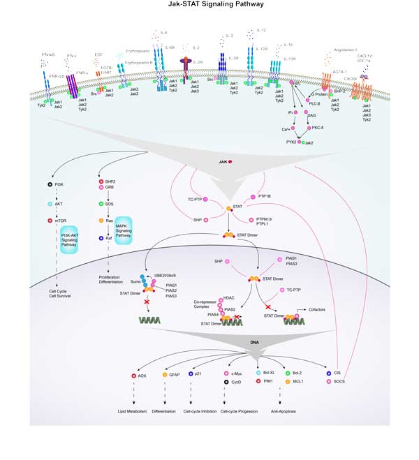
EGFR Related Signaling Pathway
EGFR is activated by binding to a variety of ligands, such as epidermal growth factor (EGF) and transforming growth factor α (TGFα), which induce the dimerization of EGFR, which in turn triggers autophosphorylation and downstream signaling. EGFR activation can activate a number of signaling pathways, including MAPK, Akt and JNK, to promote cell proliferation, differentiation, migration and survival. Abnormal activation of EGFR signaling pathway has been associated with the occurrence and development of various tumors, including non-small cell lung cancer, head and neck cancer, and breast cancer. Activation of the EGFR signaling pathway is also important for skin immunity, and EGFR ligands promote cell proliferation by activating EGFR in an autocrine form, and co-expression often predicts poor tumor prognosis.
Abnormal activation or overexpression of EGFR is closely associated with the occurrence, development, and prognosis of a variety of tumors, including non-small cell lung cancer, head and neck cancer, breast cancer, ovarian cancer, colorectal cancer, pancreatic cancer, kidney cancer, and prostate cancer. In these tumors, the mutation or overexpression of EGFR can promote the proliferation, invasion and metastasis of tumor cells, and inhibit cell apoptosis, leading to the malignant progression of tumors and the increase of treatment resistance. In addition, abnormal activation of EGFR has been implicated in some non-neoplastic diseases, such as certain skin diseases and inflammatory bowel disease.
Bioapplications of EGFR
The application of EGFR is mainly concentrated in the medical field, especially in the treatment of tumors. Egfr-targeting drugs, including small molecule tyrosine kinase inhibitors (e.g. Gefitinib, erlotinib) and monoclonal antibodies (e.g. Cetuximab, panizumab), have been developed and are widely used clinically, especially in the treatment of non-small cell lung cancer, head and neck cancer, and certain types of breast cancer. These drugs specifically inhibit the activity of EGFR, thereby slowing the proliferation and metastasis of tumor cells and improving the survival rate and quality of life of patients. In addition, the detection of EGFR is used to guide cancer treatment decisions and predict a patient's response to specific treatments.
Case Study
Case Study 1: Konstantinos Lontos, 2023
Due to several resistance mechanisms, CAR-T cell therapies remain ineffective in solid tumors. This study sought to determine whether human CAR-T cells could be enabled through a metabolic reprogramming approach. Anti-EGFR CAR-T cells were infused in NSG mice which bore A549 tumors. The tumor infiltrating lymphocytes were analyzed for exhaustion and metabolic deficiencies. Lentiviruses carrying PPAR-gamma coactivator 1α (PGC-1α), PGC-1αS571A and NT-PGC-1α constructs were used to co-transduce T cells with anti-EGFR CAR lentiviruses. Finally, researchers treated therapeutically A549-carrying NSG mice with either PGC-1α or NT-PGC-1α anti-EGFR CAR-T cells. They also analyzed the differences in the tumor-infiltrating CAR-T cells when PGC-1α is co-expressed. The results showed that transcriptomic profiling of PGC-1α-transduced CAR-T cells showed this approach effectively induced mitochondrial biogenesis, but also upregulated programs associated with effector functions. Treatment of immunodeficient animals bearing human solid tumors with these cells resulted in substantially improved in vivo efficacy.
Fig1. 107 hEGFR-targeted CARs were injected in NSG mice carrying 100 mm3 tumors.
Fig2. MitoTracker FM staining of CAR-T cells in tumor.
Case Study 2: Gui-Hua Yan, 2018
It is still beyond current reach to create protein-like specific interactions and functions on NPs by conformational engineering of nonfunctional groups on NPs. Researchers develop a conformational engineering method to create an NP-based artificial antibody, denoted "Goldbody," through conformational reconstruction of the complementary-determining regions (CDRs) of natural antibodies on gold NPs (AuNPs). The seemingly insurmountable task of controlling the conformation of the CDR loops, which are flexible and nonfunctional in the free form, was accomplished unexpectedly in a simple way. Upon anchoring both terminals of the free CDR loops on AuNPs, they managed to reconstruct the "active" conformation of the CDR loops by tuning the span between the two terminals and, as a result, the original specificity was successfully reconstructed on the AuNPs. Two Goldbodies have been created by this strategy to specifically bind with hen egg white lysozyme and epidermal growth factor receptor, with apparent affinities several orders of magnitude stronger than that of the original natural antibodies.
Fig3. Model of the anti-EGFR Goldbody in complex with sEGFR.
Fig4. SPR kinetics of the interaction between EGF and immobilized sEGFR.
EGFR involved in several pathways and played different roles in them. We selected most pathways EGFR participated on our site, such as AGE/RAGE pathway,ARMS-mediated activation,Adaptive Immune System, which may be useful for your reference. Also, other proteins which involved in the same pathway with EGFR were listed below. Creative BioMart supplied nearly all the proteins listed, you can search them on our site.
EGFR has several biochemical functions, for example, ATP binding,MAP kinase kinase kinase activity,actin filament binding. Some of the functions are cooperated with other proteins, some of the functions could acted by EGFR itself. We selected most functions EGFR had, and list some proteins which have the same functions with EGFR. You can find most of the proteins on our site.
EGFR has direct interactions with proteins and molecules. Those interactions were detected by several methods such as yeast two hybrid, co-IP, pull-down and so on. We selected proteins and molecules interacted with EGFR here. Most of them are supplied by our site. Hope this information will be useful for your research of EGFR.
Fernandez-Ulibarri, I; Hammer, K; et al. Genetic delivery of an immunoRNase by an oncolytic adenovirus enhances anticancer activity. INTERNATIONAL JOURNAL OF CANCER 136:2228-2240(2015).
Wang, YJ; Zhang, CL; et al. Investigation of phase SPR biosensor for efficient targeted drug screening with high sensitivity and stability. SENSORS AND ACTUATORS B-CHEMICAL 209:313-322(2015).


.jpg)
.jpg)