GFAP
-
Official Full Name
glial fibrillary acidic protein -
Overview
This gene encodes one of the major intermediate filament proteins of mature astrocytes. It is used as a marker to distinguish astrocytes from other glial cells during development. Mutations in this gene cause Alexander disease, a rare disorder of astrocytes in the central nervous system. Alternative splicing results in multiple transcript variants encoding distinct isoforms. [provided by RefSeq, Oct 2008] -
Synonyms
GFAP;glial fibrillary acidic protein
Recombinant Proteins
- Human
- Mouse
- Rat
- Zebrafish
- Bovine
- Porcine
- Pongo abelii
- E.coli
- HEK293
- Mammalian Cells
- Wheat Germ
- Yeast
- bovine Spinal Cord
- In Vitro Cell Free System
- Spinal Cord
- Human Brain
- Insect cells
- His
- His&SUMO
- Myc&DDK
- Non
- His&S
- His&T7
- His&GST
- Flag
- GST
- His&Myc
- His&Fc&Avi
- His&Myc&SUMO
Background

Fig1. A scheme of the different GFAP isoforms, vimentin, nestin, and synemin in mouse astrocytes. (Elly M Hol, 2015)
What is GFAP protein?
GFAP gene (glial fibrillary acidic protein) is a protein coding gene which situated on the long arm of chromosome 17 at locus 17q21. GFAP is an intermediate filament protein that is predominantly found in astrocytes within the central nervous system (CNS), as well as in non-myelinating Schwann cells in the peripheral nervous system (PNS) and enteric glial cells. It serves as a key component of the cytoskeleton in these cells and is involved in maintaining their mechanical strength and structural integrity. The GFAP protein is consisted of 432 amino acids and GFAP molecular weight is approximately 49.9 kDa.
What is the function of GFAP protein?
The GFAP protein is a key intermediate filament protein primarily expressed in astrocytes, which are star-shaped glial cells in the central nervous system. Its primary function is to provide structural support and maintain the integrity of astrocyte processes, thereby contributing to the stability of the cellular cytoskeleton. GFAP also plays a crucial role in maintaining the blood-brain barrier, facilitating cell-cell interactions, and participating in the response to brain injuries or neurodegenerative diseases by promoting scar formation and modulating immune responses. Dysregulation of GFAP expression is often associated with various neurological disorders, including Alzheimer's disease, multiple sclerosis, and Alexander disease, underscoring its importance in neural health and disease.
GFAP related signaling pathway
The GFAP (Glial Fibrillary Acidic Protein) is primarily involved in the structural integrity and function of astrocytes, crucial glial cells in the central nervous system. While GFAP itself does not directly participate in signaling pathways, its expression and degradation are influenced by various signaling mechanisms. For instance, during brain injury or neurodegenerative diseases, signaling pathways such as MAPK/ERK and PI3K/AKT can modulate GFAP expression, promoting astrogliosis and scar formation. Additionally, inflammatory cytokines like TNF-α and IL-1β can activate JAK/STAT pathways, leading to increased GFAP production. These pathways highlight the role of GFAP in the response to CNS injuries and pathologies, emphasizing its importance in maintaining neural homeostasis and repair processes.
GFAP related diseases
One of the most significant GFAP-related diseases is autoimmune glial fibrillary acidic protein astrocytopathy (GFAP-A), which is a treatable central nervous system autoimmune inflammatory disease characterized by meningeal, brain, spinal cord, and optic nerve involvement. GFAP-A can manifest with a variety of neurological symptoms, including encephalitis, myelitis, and raised intracranial pressure. Magnetic resonance imaging (MRI) may show specific changes like linear or radial vascular patterns enhancing around the ventricles, which can be a characteristic imaging feature of GFAP-A. Additionally, GFAP-A has been associated with certain types of cancer, particularly ovarian teratomas, suggesting a potential paraneoplastic origin in some cases.
Bioapplications of GFAP
GFAP has several bioapplications, particularly in the field of neuroscience and diagnostics. It serves as a biomarker for various neurological disorders, including Alzheimer's disease, multiple sclerosis, and traumatic brain injury, aiding in early diagnosis and monitoring disease progression. Additionally, GFAP is involved in the development of therapeutic strategies for neurodegenerative diseases, where modulating its expression can influence astrocyte behavior and neural repair processes. Moreover, GFAP-targeted imaging techniques are employed to visualize astrocyte activation and glial scar formation, providing insights into CNS pathology and guiding intervention strategies. These applications underscore GFAP's significance in understanding and treating neurological conditions.
Case Study
Case Study 1: Ai-Wen Yang, 2022
Alexander disease is a genetic astrocyte disorder caused by dominant GFAP gene mutations. The mechanism by which these mutations cause brain degeneration is not well understood. Researchers studied 14 GFAP missense mutations in the rod domain's impact on filament assembly in vitro. Internal rod mutants formed shorter filaments, while rod end mutants created atypical structures with short lengths, irregular widths, and aggregations. When introduced into primary astrocytes, these GFAP mutants with assembly defects often resulted in cytoplasmic aggregates that were resistant to extraction. This resistance was also found in Alexander disease patient brain tissues, where insoluble GFAP accumulated in Rosenthal fibers.

Fig1. Electron micrographs of in vitro assembly structures generated by WT and mutant GFAPs.

Fig2. Cells transduced with the empty vector (lane 1) or the indicated GFAP constructs (lanes 2–6) were extracted, and the resulting supernatant (S) and pellet (P) fractions were analyzed.
Case Study 2: Rebeca Uceda-Castro, 2022
Glioma is the most prevalent malignant primary brain tumor in adults, and its highly invasive nature makes it incurable, highlighting the need to understand the mechanisms behind its invasion. GFAP, a protein specific to astrocyte- and neural stem cell-derived gliomas, undergoes alternative splicing changes in malignancy, with the GFAPα form decreasing in higher-grade tumors and the GFAPδ form becoming more dominant. Using intravital imaging and an ex vivo brain slice model, we found that GFAPδ and GFAPα isoforms differently regulate glioma cell dynamics. Depletion of either isoform enhances the migration of glioma cells, with GFAPδ-depleted cells migrating randomly and GFAPα-depleted cells showing a persistent, directional invasion into the brain. This study reveals that the GFAP network's composition influences the migratory behavior of glioma cells.

Fig3. Protein levels of GFAPδ and all GFAP isoforms (GFAPpan) in the 12 different cell clones.

Fig4. Quantification of tumour density for each indicated tumour type.
Quality Guarantee
High Purity
.jpg)
Fig1. SDS-PAGE (GFAP-885H)
.
.jpg)
Fig2. SDS-PAGE (GFAP-2745H)
Involved Pathway
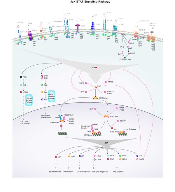
GFAP involved in several pathways and played different roles in them. We selected most pathways GFAP participated on our site, such as Jak-STAT signaling pathway, which may be useful for your reference. Also, other proteins which involved in the same pathway with GFAP were listed below. Creative BioMart supplied nearly all the proteins listed, you can search them on our site.
| Pathway Name | Pathway Related Protein |
|---|---|
| Jak-STAT signaling pathway | IL11RA2,IL7R,IL2RB,IL4,MCL1,IL29,IFNA16,LEPA,SOCS1A,IL12RB2 |
Protein Function
GFAP has several biochemical functions, for example, integrin binding,kinase binding,protein binding. Some of the functions are cooperated with other proteins, some of the functions could acted by GFAP itself. We selected most functions GFAP had, and list some proteins which have the same functions with GFAP. You can find most of the proteins on our site.
| Function | Related Protein |
|---|---|
| protein binding | ATPAF2,NIPA2,PADI4,CXXC11,DCPS,BTBD1,GABPB2,AKR1A1,RAB24,SDC1 |
| structural constituent of cytoskeleton | SPTAN1,TUBGCP4,SYNM,LOR,TUBB5,TUBG1,TLN2A,MAPT,KRT5,KRT14 |
| kinase binding | EMP2,SLC2A1,STRADA,DLG3,HAP1,JAKMIP3,BCL10,PPP1R9B,PTPN22,CTNNB1 |
| integrin binding | EMP2,ICAM5,Tcam1,SYK,Npnt,NOV,L1CAM,ICAM4,COL5A1,DST |
Interacting Protein
GFAP has direct interactions with proteins and molecules. Those interactions were detected by several methods such as yeast two hybrid, co-IP, pull-down and so on. We selected proteins and molecules interacted with GFAP here. Most of them are supplied by our site. Hope this information will be useful for your research of GFAP.
SH3YL1;KRT13;PIAS2;MOS;TRIM27;KRT15
GFAP Related Signal Pathway
Resources
Research Area
NeuroinflammationNeural Stem Cell Markers
Glial Lineage Markers
Intermediate Filaments
Neural Progenitor Cell Markers
Glioma Biomarkers
Related Services
Related Products
References
- Sun, Y; Lehmbecker, A; et al. STAT3 represents a molecular switch possibly inducing astroglial instead of oligodendroglial differentiation of oligodendroglial progenitor cells in Theiler's murine encephalomyelitis. NEUROPATHOLOGY AND APPLIED NEUROBIOLOGY 41:347-370(2015).

