EPCAM
-
Official Full Name
epithelial cell adhesion molecule -
Overview
This gene encodes a carcinoma-associated antigen and is a member of a family that includes at least two type I membrane proteins. This antigen is expressed on most normal epithelial cells and gastrointestinal carcinomas and functions as a homotypic calcium-independent cell adhesion molecule. The antigen is being used as a target for immunotherapy treatment of human carcinomas. Mutations in this gene result in congenital tufting enteropathy. -
Synonyms
EPCAM;epithelial cell adhesion molecule;ESA;KSA;M4S1;MK-1;DIAR5;EGP-2;EGP40;KS1/4;MIC18;TROP1;EGP314;HNPCC8;TACSTD1;antigen identified by monoclonal antibody AUA1;tumor-associated calcium signal transducer 1;17-1A;323/A3;CD326;CO-17A;EGP34;EGP40;GA733-2;HEA125;Ly74;MH99;MOC31;TACST-1;Adenocarcinoma-associated antigen;Cell surface glycoprotein Trop-1;Epithelial cell surface antigen;Epithelial glycoprotein;Epithelial glycoprotein 314;KS 1/4 antigen;Major gastrointestinal tumor-associated protein GA733-2;TACSTD1;human epithelial glycoprotein-2;Ep-CAM;membrane component, chromosome 4, surface marker (35kD glycoprotein);EGP;M1S2;CD326 antigen;Hegp314;ACSTD1;Adecatumumab;503605-66-1;Citatuzumab bogatox;945228-49-9;Citatuzumab bogatox;945228-49-9;Tucotuzumab celmoleukin;339986-90-2;Solitomab;503605-66-1
Recombinant Proteins
- Mouse
- Human
- Monkey
- Rat
- Rhesus macaque
- Cynomolgus
- Chicken
- Zebrafish
- Pig
- Mammalian cells
- HEK293
- Human Cell
- HEK293 cells
- Mammalian Cell
- Sf9 Insect Cell
- Wheat Germ
- Human cells
- E.coli
- Insect Cell
- Mouse
- HEK293T
- Mammalian cell
- In Vitro Cell Free System
- E.coli expression system
- Fc
- His
- Flag
- Fc&Avi
- Non
- His&Flag&StrepII
- GST
- His&T7
- His&Avi
- His&Fc&Avi
- His&GST
- Strep
- Myc&DDK
- His&SUMO
- mFc
Background
What is EPCAM Protein?
EPCAM gene (epithelial cell adhesion molecule) is a protein coding gene which situated on the short arm of chromosome 2 at locus 2p21. This gene encodes a carcinoma-associated antigen and is a member of a family that includes at least two type I membrane proteins. This antigen is expressed on most normal epithelial cells and gastrointestinal carcinomas and functions as a homotypic calcium-independent cell adhesion molecule. The antigen is being used as a target for immunotherapy treatment of human carcinomas. The EPCAM protein is consisted of 314 amino acids and EPCAM molecular weight is approximately 34.9 kDa.
What is the Function of EPCAM Protein?
EPCAM is a cell surface glycoprotein that plays a role in a variety of physiological and pathological processes, and it was originally discovered as a tumor antigen. EPCAM is involved in the regulation of intercellular adhesion strength and tissue plasticity, and influences cell proliferation and differentiation. In tissue regeneration and stem cell research, EPCAM helps maintain the stem cell properties of cells. At the molecular level, EPCAM is involved in homologous adhesion between cells through its extracellular domains, while its intracellular domains interact with the cytoskeleton to influence cell morphology and movement. In addition, EPCAM can produce active extracellular and intracellular fragments through controlled endometrial proteolysis, which in turn affects a variety of signal transduction processes including WNT and Ras/Raf signaling pathways.
EPCAM Related Signaling Pathway
The extracellular domain (EpEX) of EpCAM binds to the epidermal growth factor receptor (EGFR) and activates the AKT and MAPK signaling pathways. This can inhibit the function of the transcription factor FOXO3a and stabilize the PD-L1 protein, thus promoting tumor progression. EpCAM produces active extracellular and intracellular fragments through controlled intimal proteolysis (RIP). In particular, the intracellular fragment of EpCAM (EpICD) can be transported to the nucleus, form complexes with β-catenin and Lef-1, promote gene transcription, and is associated with the proliferation of tumor cells. EpCAM regulates Wnt signaling through RIP and influences cell proliferation and cell fate determination. EpCAM participates in intercellular adhesion and influences intercellular interactions by forming autodimers or heterodimers.
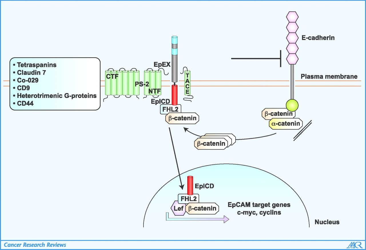
Fig1. Schematic of signaling pathways of EpCAM. (Markus Munz, 2009)
EPCAM Related Diseases
EPCAM is a glycoprotein expressed in a variety of epithelial cell and epithelial-derived tumors, and its abnormal expression has been associated with a variety of diseases. It is involved in regulating cell adhesion, proliferation, differentiation, migration and signaling. Studies have shown that EPCAM is abnormally expressed in many human tumors and that its expression levels correlate with disease prognosis, particularly in colorectal, gastric, prostate, and breast cancers. In addition, the role of EPCAM signaling in tumor progression is multifaceted, including promoting tumor cell invasion, immune escape, and activation of downstream oncogenes.
Bioapplications of EPCAM
EPCAM is a known tumor antigen on epithelial tumors, circulating tumor cells and tumor stem cells, and monoclonal antibodies against EPCAM have been used in a variety of cancer therapies. Epcam-car T cell therapy, which uses chimeric antigen receptor modified T cells to target EpCAM, has been shown to be safe and effective in solid tumors in preclinical and clinical studies. As a prognostic marker, therapeutic target and anchor molecule of CTC, EpCAM plays an important role in the detection and analysis of CTC. Antibody drug couplings (ADCs), such as Oportuzumab Monatox and Citatuzumab Bogatox, use EpCAM antibodies to bind to toxins to target cancer cells for precision killing
Case Study
Case Study 1: Sushree Shankar Panda, 2024
Once in the nucleus, EpICD transcriptionally regulates EpCAM target genes that; however, remains unclear whether Wnt signaling is modulated by EpICD activity. Patient-derived organoids (PDOs), patient-derived xenografts (PDXs), and various CRC cell lines were used to study the roles of EpCAM and EpICD in Wnt receptor expression. EpCAM signaling was intervened with our humanized form of EpCAM neutralizing antibody, hEpAb2-6. Functional assays including in vivo tumor formation, organoid formation, spheroid and colony formation experiments were performed to study Wnt related phenomena. The results showed that EpICD interacted with the promoters of Wnt receptors (FZD6 and LRP5/6) thus upregulated their transcriptional activity inducing Wnt signaling. Furthermore, activation of Wnt-pathway-associated kinases in the β-Catenin destruction complex (GSK3β and CK1) induced γ-secretase activity to augment EpICD shedding, establishing a positive-feedback loop.

Fig1. IFS for hEpAb2-6 binding to EpCAM in PDOs.
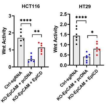
Fig2. Wnt activity as detected by TOP flash reporter assay in indicated control and EpCAM knockout cell lines with or without EpICD plasmid transfection.
Case Study 2: Dandan Zhang, 2020
Epithelial cell adhesion molecule (EpCAM), which is a transmembrane glycoprotein, is related to tumor progression. Researchers demonstrated that EpCAM plays important roles in proliferation, apoptosis, and metastasis during breast cancer (BC) progression. But the role of N-glycosylation in EpCAM in tumor aggressiveness is not clear. Here, they evaluated the role of N-glycosylation of EpCAM in stemness and epithelial-mesenchymal transition (EMT) characteristics. EpCAM overexpression increases the expression of stemness markers (NANOG,SOX2, and OCT4) and EMT markers (N-cadherin and vimentin) under the condition of hypoxia in BC. Knockdown of EpCAM and mutation of N-glycosylation of EpCAM maintained in severe hypoxia lead to a significant reduction of stemness/EMT markers. In addition, N-glycosylation of EpCAM is a crucial factor during this process. This demonstrates that EpCAM has a novel regulatory role in stemness/EMT dependence of hypoxia-inducible factor 1-alpha via regulating nuclear factor kappa B in BC cells.
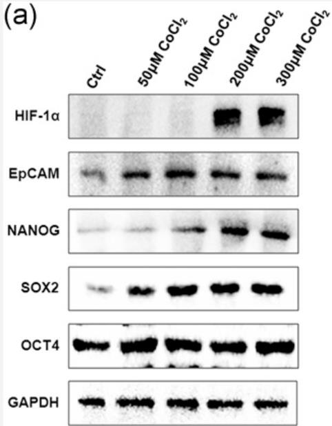
Fig3. CoCl2 upregulated the expression of HIF-1α, EpCAM, and stem cell markers.
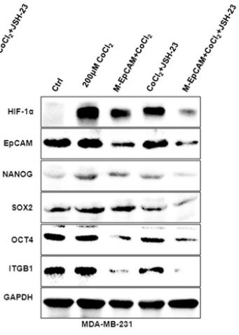
Fig4. Cell lysates were determined with the antibodies of HIF-1α, EpCAM, NANOG, SOX2, and OCT4 by western blot.
Quality Guarantee
High Purity
.jpg)
Fig1. SDS-PAGE (EPCAM-051H)
.
.jpg)
Fig2. SDS-PAGE (EPCAM-7817H)
Involved Pathway
EPCAM involved in several pathways and played different roles in them. We selected most pathways EPCAM participated on our site, such as , which may be useful for your reference. Also, other proteins which involved in the same pathway with EPCAM were listed below. Creative BioMart supplied nearly all the proteins listed, you can search them on our site.
| Pathway Name | Pathway Related Protein |
|---|
Protein Function
EPCAM has several biochemical functions, for example, cadherin binding involved in cell-cell adhesion,protein binding,protein complex binding. Some of the functions are cooperated with other proteins, some of the functions could acted by EPCAM itself. We selected most functions EPCAM had, and list some proteins which have the same functions with EPCAM. You can find most of the proteins on our site.
| Function | Related Protein |
|---|---|
| protein binding | KDM1A,OLFM4,TH1L,ANKHD1,SYT1,ADRBK1,HBQ1,SEMA7A,SMC1A,SSB |
| protein complex binding | WASF2,ABI1,NOG,SKAP1,CAPG,TUB,MYC,FLCN,NR0B2,FGFR1 |
Interacting Protein
EPCAM has direct interactions with proteins and molecules. Those interactions were detected by several methods such as yeast two hybrid, co-IP, pull-down and so on. We selected proteins and molecules interacted with EPCAM here. Most of them are supplied by our site. Hope this information will be useful for your research of EPCAM.
CFTR
Resources
Related Services
Related Products
References
- Hyun, KA; Lee, TY; et al. Two-stage microfluidic chip for selective isolation of circulating tumor cells (CTCs). BIOSENSORS & BIOELECTRONICS 67:86-92(2015).
- Gu, YJ; Ju, C; et al. Detection of circulating tumor cells in prostate cancer based on carboxylated graphene oxide modified light addressable potentiometric sensor. BIOSENSORS & BIOELECTRONICS 66:24-31(2015).

