Yellow Fluorescent Protein
| Cat.No. : | YFP-101 |
| Product Overview : | Fluorescent protein ideal for subcellular labeling/visualization. The recombinant YFP (Yellow Fluorescent Protein) is expressed and purified from transformed E. coli using a method that ensures high purity and maximal YFP fluorescence. Endotoxin has been removed, so the protein is suitable for in vivo injection or cell culture applications. The protein is a 26.4 kDa monomer with 238 amino acids, Ex./Em. = 525/538 nm, extinction coefficient 27390. |
- Specification
- Gene Information
- Related Products
- Case Study
- Application
- Download
| Source : | E.coli |
| Tag : | Non |
| Description : | Yellow fluorescent protein (YFP) is a genetic mutant of green fluorescent protein (GFP) originally derived from the jellyfish Aequorea victoria. Its excitation peak is 514 nm and its emission peak is 527 nm. Like the parent GFP, YFP is a useful tool in cell and molecular biology thanks to its properties useful for fluorescence microscopy. Three improved versions of YFP are Citrine, Venus, and Ypet. They have reduced chloride sensitivity, faster maturation, and increased brightness (defined as the product of the extinction coefficient and quantum yield). Typically, YFP serves as the acceptor for genetically-encoded FRET sensors of which the most likely donor FP is monomeric cyan fluorescent protein (mCFP). The red-shift relative to GFP is caused by a Pi-Pi stacking interaction as a result of the T203Y substitution introduced by mutation, which essentially increases the polarizability of the local chromophore environment as well as providing additional electron density into the chromophore. |
| Form : | Lyophilized protein |
| Molecular Mass : | 26.4 kDa |
| Endotoxin : | <0.1 ng/μg |
| Purity : | ≥97%. |
| Usage : | For Research Use Only! Not to be used in humans. |
| Storage : | At -20 centigrade. |
| Reconstitution : | Reconstitute with dH2O to 1 mg/mL. |
| Handling : | Centrifuge the vial prior to opening. |
| Synonyms | YFP; Yellow Fluorescent Protein |
| ◆ Recombinant Proteins | ||
| YFP-149 | Recombinant Yellow Fluorescent Protein, His-tagged | +Inquiry |
| YFP-10 | Recombinant Yellow Fluorescent Protein | +Inquiry |
| YFP-01 | Recombinant Yellow Fluorescent Protein | +Inquiry |
| YFP-02 | Recombinant YFP Protein | +Inquiry |
| YFP-007E | Recombinant Yellow Fluorescent Protein | +Inquiry |
| ◆ Native Proteins | ||
| YFP-101 | Yellow Fluorescent Protein | +Inquiry |
Case 1: Jusuk I, et al. Sci Rep. 2015
Photostability is essential for super-resolution imaging using fluorophores. enhanced yellow fluorescent protein (eYFP) is popular in cell biology for its quick blinking and UV-induced photoactivation. Researchers developed an assay to study single eYFP photostabilization, showing a 6-fold improvement under oxygen-free conditions and with aliphatic thiols. By attaching eYFP to single-stranded DNA on a DNA origami structure in solution, they conducted single-molecule photophysical studies under near-physiological conditions. The improved properties were demonstrated for super-resolution microscopy, using DNA origami as nanorulers and imaging microtubules in fixed Vero cells.
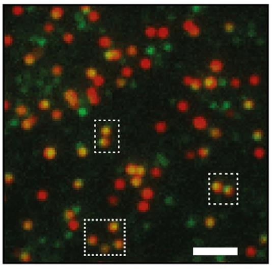
Fig1. Corresponding TIRF image of overlaid channels of eYFP-emission (green) and ATTO647N-emission (red).
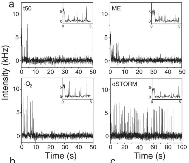
Fig2. Representative fluorescence transients of eYFP in four different buffer conditions.
Case 2: Bierig T, et al. Front Bioeng Biotechnol. 2020
The 2019-nCoV, responsible for the COVID-19 pandemic, requires detailed study to inform vaccine and diagnostic assay development. Researchers created a protocol for expressing and purifying a yellow fluorescent protein (YFP)-tagged 2019-nCoV spike receptor-binding domain (RBD). This construct in pcDNA 4/TO includes an IFNα2 signal peptide, eYFP, FLAG-tag, HRV3C cleavage site, 2019-nCoV RBD, and an 8x His-tag. After stable transfection into HEK 293 cells, the fusion protein was secreted and purified using Ni-NTA chromatography, yielding a soluble, monodisperse protein confirmed by SEC and electron microscopy. The presence of N-linked glycosylations was verified, and complex formation with human ACE2's peptidase domain was demonstrated. The fusion protein has potential applications in cell binding studies, fluorescent labeling of virus-binding sites, and the development of immunization and detection assays.
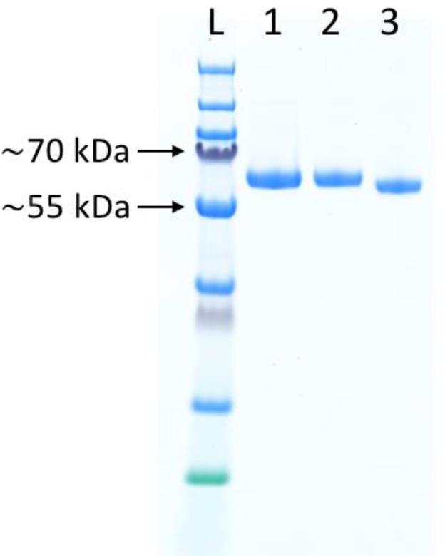
Fig1. SDS-PAGE analysis of the YFP-S_RBD fusion protein.
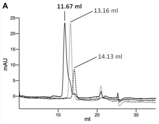
Fig2. Analysis of complex formation by analytical SEC.
Yellow Fluorescent Protein (YFP) is a widely used fluorescent marker that belongs to a mutant of green fluorescent protein (GFP). YFP's excitation peak is usually around 514nm and its emission peak is around 527nm, which makes it glow bright yellow. Such spectral characteristics make YFP particularly suitable for multi-color fluorescence labeling and imaging, and it can be used in combination with other colored fluorescent proteins (such as ECFP, EYFP, etc.) to achieve multi-label experiments.
In biological research, YFP is often used as a genetic marker to study protein localization, interaction, and dynamic processes in vivo and in vitro. Due to its high brightness and good light stability, YFP is particularly suitable for time series analysis and fluorescence resonance energy transfer (FRET) experiments. In FRET technology, YFP can be used as an energy receptor in combination with another fluorescent protein, such as CFP.
YFP can be used to develop biosensors that detect changes in specific ion concentrations or pH levels within cells. During drug development, YFP is used as a reporter gene to monitor the effect of a drug on cell function. YFP and its variants are used in super-resolution imaging to help reveal details of cell structure and molecular assembly. With the development of molecular biology techniques, many improved variants of YFP have been derived, which have higher brightness, better light stability or different spectral characteristics to meet specific experimental needs.
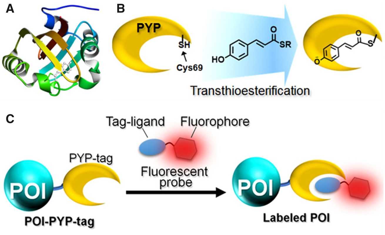
Fig1. The photoactive yellow protein (PYP)-based labelling system. (Naresh Kumar, 2019)
Not For Human Consumption!
Inquiry
- Reviews
- Q&As


Ask a Question for All YFP Products
Required fields are marked with *
My Review for All YFP Products
Required fields are marked with *
Inquiry Basket