Active Recombinant Cynomolgus PDGFB protein(Ser82-Thr190), mFc-tagged
| Cat.No. : | PDGFB-209C |
| Product Overview : | Recombinant Cynomolgus PDGFB (EHH65851.1) (Ser82-Thr190) was expressed in HEK293 with the Fc region of Mouse IgG1 at the N-terminus. |
- Specification
- Gene Information
- Related Products
- Case Study
- Application
- Download
| Species : | Cynomolgus |
| Source : | HEK293 |
| Tag : | mFc |
| Protein Length : | Ser82-Thr190 |
| Form : | Lyophilized from sterile PBS, pH 7.4. Normally 5 % - 8 % trehalose, mannitol and 0.01% Tween80 are added as protectants before lyophilization. |
| Bio-activity : | 1. Measured in a cell proliferation assay using Balb/c 3T3 mouse embryonic fibroblasts. The ED50 for this effect is typically 5-25 ng/ml. 2. Measured by its binding ability in a functional ELISA. Immobilized Cynomolgus PDGFRB-His at 10 μg/ml (100 μl/well) can bind Cynomolgus S4-mFc3-PDGFB, The EC50 of Cynomolgus S4-mFc3-PDGFB is 3.6-7.4 ng/ml. |
| Molecular Mass : | The recombinant cynomolgus PDGFB is a disulfide-linked homodimer. The reduced monomer comprises 345 amino acids and has a calculated molecular mass of 38.9 KDa. The apparent molecular mass of it is approximately 43 and 32 KDa in SDS-PAGE. |
| Endotoxin : | < 1.0 EU per μg of the protein as determined by the LAL method. |
| Purity : | > 95 % as determined by SDS-PAGE |
| Storage : | Samples are stable for up to twelve months from date of receipt at -20°C to -80°C. Store it under sterile conditions at -20°C to -80°C. It is recommended that the protein be aliquoted for optimal storage. Avoid repeated freeze-thaw cycles. |
| Reconstitution : | It is recommended that sterile water be added to the vial to prepare a stock solution of 0.2 ug/ul. Centrifuge the vial at 4°C before opening to recover the entire contents. |
| ◆ Recombinant Proteins | ||
| PDGFB-5843C | Recombinant Chicken PDGFB | +Inquiry |
| PDGFB-2571H | Recombinant Human PDGFB Protein, His-tagged | +Inquiry |
| PDGFB-209C | Active Recombinant Cynomolgus PDGFB protein(Ser82-Thr190), mFc-tagged | +Inquiry |
| Pdgfb-316P | Active Recombinant Mouse Pdgfbb Protein (110 aa) | +Inquiry |
| PDGFB-1174H | Recombinant Human PDGFB Protein, His-tagged | +Inquiry |
| ◆ Cell & Tissue Lysates | ||
| PDGFB-1321HCL | Recombinant Human PDGFB cell lysate | +Inquiry |
Case 1: Contreras O, et al. Cell Signal. 2021
PDGFs influence development, tissue repair, and healing by binding to PDGFRα and PDGFRβ. Despite their importance, the impact of PDGF signaling on muscle development and regeneration is not well understood. Researchers investigated PDGF-PDGFR gene expression in skeletal muscle myogenesis and found that myogenic cells differentially express PDGF ligands and receptors during this process. Adult muscle stem cells and myoblasts mainly express PDGFRβ. Muscle stem cell activation changes PDGF gene expression, and PDGF-AB and PDGF-BB in myoblasts activate RAS-ERK1/2 and PI3K-AKT pathways, promoting chemotaxis and mitogenesis. The PDGFR inhibitor AG1296 blocks these activations, inhibiting myoblast migration, proliferation, and cell cycle progression. AG1296 also arrests myoblast cell cycles in G0/G1. PDGF-AA did not significantly affect these processes. Additionally, myogenic differentiation reduces PDGFRα and PDGFRβ expression, and forced PDGFRα expression hinders myogenesis.
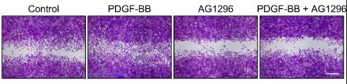
Fig1. Representative images of C2C12 myoblasts control-treated or treated with PDGF-BB and/or AG1296.
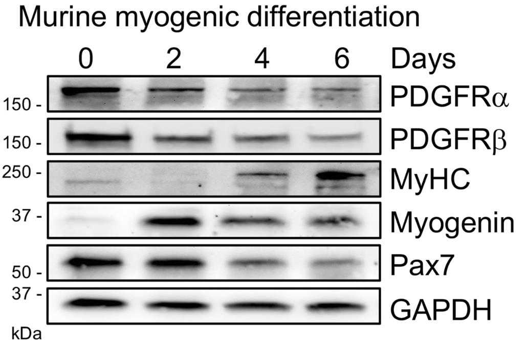
Fig2. PDGFRα, PDGFRβ, MyHC, myogenin, Pax7, and GAPDH levels were analyzed by western blot.
Case 2: Zhang Y, et al. Cancers (Basel). 2022
Platelets are a significant source of PDGFB and are often activated in tumors, releasing growth factors. To study PDGFB's role in tumors, we developed a mouse model with platelet-specific PDGFB knockout (pl-PDGFB KO). This resulted in a 10-fold PDGFB reduction in the tumor environment, fewer cancer-associated fibroblasts, and less ECM deposition, specifically fibronectin and collagen I, in a pancreatic neuroendocrine cancer model. Tumors from pl-PDGFB KO mice showed reduced myosin light chain phosphorylation, which affects cell contraction and TGFβ release. Correspondingly, TGFβ signaling, as indicated by phosphorylated Smad2, was impaired, explaining the decreased ECM deposition.
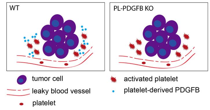
Fig1. PDGFB is released from activated platelets in the TME in WT mice, but not in pl-PDGFB KO mice.
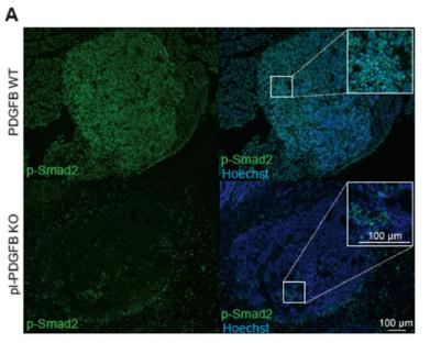
Fig2. Tumors from 14-week old WT and pl-PDGFB KO RT2-positive mice were immunostained for p-Smad2.
Recombinant Cynomolgus PDGFB protein plays a significant role in various medical applications, particularly in muscle regeneration and repair, hematological treatments, and cancer therapy. In muscle regeneration, PDGFB is crucial for regulating the behavior and fate of muscle stem cells, enhancing muscle maintenance and recovery post-injury. It aids in the proliferation and recruitment of mesenchymal stem cells, improving the implantation and self-renewing capability of hematopoietic cells, which is beneficial for treating blood disorders. In cancer therapy, particularly for glioblastoma, PDGFB influences tumor growth and metastasis by engaging in diverse cellular processes and is proposed as a potential therapeutic target. Its role extends to modulating the tumor microenvironment, affecting the deposition of extracellular matrix and signal transduction pathways which are critical for tumor progression and prognosis.
In scientific research, PDGFB serves as an essential tool in cell biology studies involving cell proliferation, migration, differentiation, and cycle progression, notably in muscle and stem cell research. It is employed in modeling diseases like muscle disorders and tumors using 3D cell cultures, aiding in drug discovery processes. The protein is pivotal in signal transduction studies, especially within the PDGF-PDGFR pathways relevant to muscle regeneration and disease conditions. Industrially, PDGFB finds its role in biopharmaceuticals for drug preparation aimed at wound healing and tissue repair, and as an additive in cell culture systems to promote cell expansion and differentiation. Additionally, it serves as a diagnostic tool where its expression levels can act as biomarkers in tumor diagnosis, aiding in evaluating prognosis and therapeutic responses.
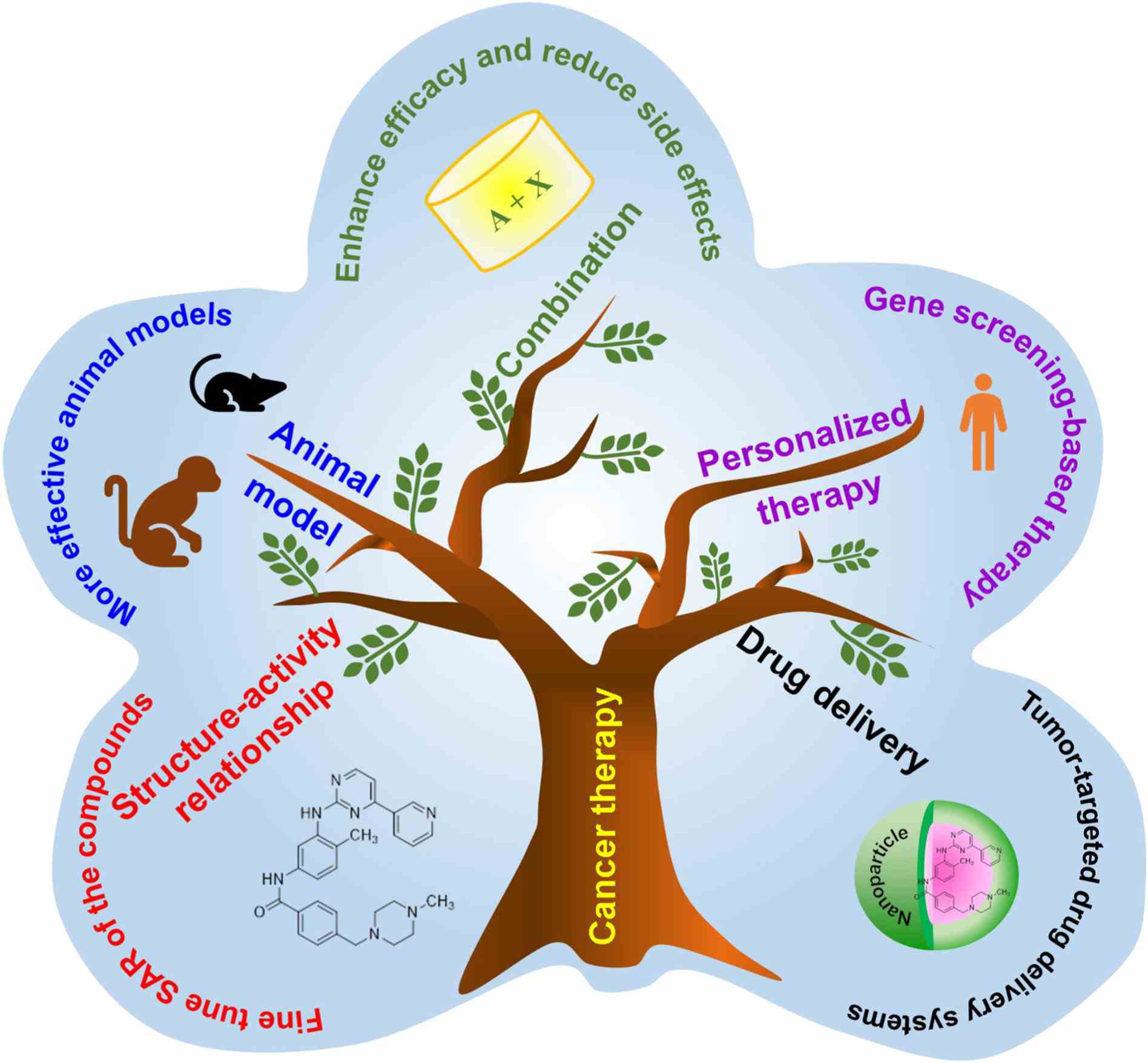
Fig1. The future directions for cancer therapy by targeting the PDGF/PDGFR signaling pathway. (Xiang Zou, 2022)
Not For Human Consumption!
Inquiry
- Reviews
- Q&As


Ask a Question for All PDGFB Products
Required fields are marked with *
My Review for All PDGFB Products
Required fields are marked with *
Inquiry Basket