Recombinant Mouse HMMR Protein
| Cat.No. : | HMMR-7742M |
| Product Overview : | Recombinant Mouse HMMR full length or partial length protein was expressed. |
- Specification
- Gene Information
- Related Products
- Case Study
- Application
- Download
| Species : | Mouse |
| Source : | Mammalian Cells |
| Tag : | His |
| Form : | Liquid or lyophilized powder |
| Endotoxin : | < 1.0 EU per μg of the protein as determined by the LAL method. |
| Purity : | >80% |
| Notes : | This item requires custom production and lead time is between 5-9 weeks. We can custom produce according to your specifications. |
| Storage : | Store it at +4 ºC for short term. For long term storage, store it at -20 ºC~-80 ºC. |
| Storage Buffer : | PBS buffer |
| Gene Name | Hmmr hyaluronan mediated motility receptor (RHAMM) [ Mus musculus ] |
| Official Symbol | HMMR |
| Gene ID | 15366 |
| mRNA Refseq | NM_013552.2 |
| Protein Refseq | NP_038580.2 |
| MIM | |
| UniProt ID | Q00547 |
| ◆ Recombinant Proteins | ||
| HMMR-195H | Recombinant Human HMMR Protein, His-tagged | +Inquiry |
| HMMR-1286HFL | Recombinant Full Length Human HMMR Protein, C-Flag-tagged | +Inquiry |
| HMMR-2776H | Recombinant Human HMMR Protein (Met1-Lys651), C-His tagged | +Inquiry |
| HMMR-1082H | Recombinant Human HMMR Protein, His (Fc)-Avi-tagged | +Inquiry |
| HMMR-4202H | Recombinant Human HMMR Protein, Myc/DDK-tagged, C13 and N15-labeled | +Inquiry |
| ◆ Cell & Tissue Lysates | ||
| HMMR-5468HCL | Recombinant Human HMMR 293 Cell Lysate | +Inquiry |
| HMMR-5469HCL | Recombinant Human HMMR 293 Cell Lysate | +Inquiry |
Case 1: Chen YJ, et al. Elife. 2024
Microtubule cytoskeleton plays a variety of important roles in neuronal morphogenesis. Although many microtubule-associated proteins (MAPs) have been shown to be involved in neuronal morphogenesis, the functions of many more remain to be determined. This study focused on a mouse MAP called HMMR, which was initially identified as a hyaluronic acid-binding protein and was later found to also have the ability to bind microtubules and centrosomes. The content of HMMR is very high in the microtubules of neurons, and changing the level of HMMR significantly affects the morphology of neurons. Unlike cells during mitosis that are confined to the centrosomes, HMMR is localized to microtubules along axons and dendrites. In addition, the transient expression of HMMR enhanced the stability of neuronal microtubules and increased the formation frequency of growing microtubules on neurites. HMMR regulates the microtubule localization of non-centrosomal microtubule nucleoforming protein TPX2 on neurites, providing an explanation for how HMMR promotes microtubule growth.
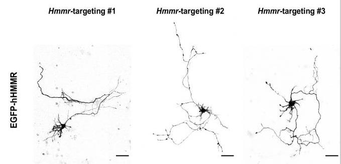
Fig1. Representative EGFP images of mouse hippocampal neurons co-transfected with EGFP-hHMMR.
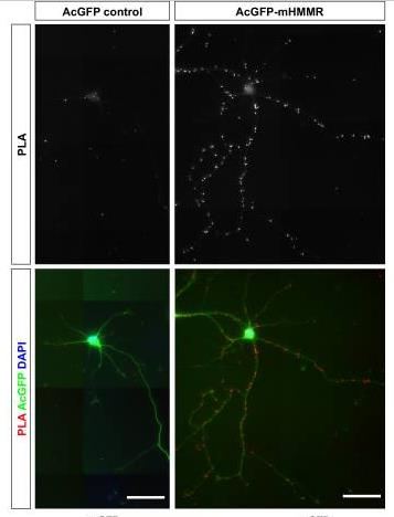
Fig2. PLA images of AcGFP control (upper left) or AcGFP-mHMMR (upper right) and β-III-tubulin in 7 DIV dissociated hippocampal neurons.
Case 2: Tolg C, et al. Am J Pathol. 2012
Hyaluronic acid regulates inflammation and fibroplasia through its activation during damage and repair of diseases such as cancer. Hyaluronic acid-binding peptides have been identified that can alter fiber production during skin wound repair. These peptides were screened from phage display libraries of 7 to 15 amino acids by binding to hyaluronic acid-agarose beads, and their ability to block fibroblast responses to hyaluronic acid oligosaccharides (10 kDa) was tested. A 15-amino acid peptide (P15-1), homologous to the hyaluronic acid-binding sequence of the hyaluronic acid-mediated motor receptor (RHAMM), is the most potent inhibitor. P15-1 has a binding affinity of K(d) = 10^(-7) with 10 kDa hyaluronic acid, and particularly mimics RHAMM because it significantly reduces the binding of hyaluronic acid oligosaccharides to recombinant RHAMM, while having no effect on recombinant CD44 or TLR2,4. And altered wound repair in wild-type but not RHAMM(-/-) mice. A local application of P15-1 to full-layer resected rat wounds significantly reduced wound macrophage count, fibroblast count, and blood vessel density compared to a mixed, negative control peptide. Wound collagen 1, transforming growth factor β-1, and alpha-smooth muscle actin decreased, while Tenascin C increased, suggesting that P15-1 promoted a form of scar-free healing. Signal/microarray analysis revealed that P15-1 blocked the Rhamm-regulated focused adhesion kinase pathway in fibroblasts.
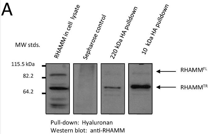
Fig1. RHAMM binds to 220-kDa and 10-kDa HA.
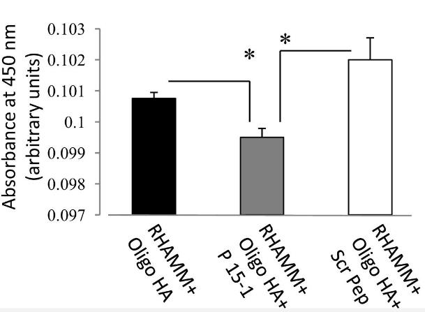
Fig2. ELISA plates were coated with recombinant RHAMM followed by incubation with HA oligosaccharides (10 kDa) together with P15-1 or Scr Pep.
Murine Hmmr proteins (hyaluronic acid-mediated motoreceptors) are a class of proteins with predicted hyaluronic acid-binding activity, mainly located in centrosomes and cytoplasm. Hmmr protein is of great significance in the study of diseases such as cancer, inflammation and aging, and can be used to study cell movement, cell cycle regulation, and intracellular signaling.
Hmmr proteins are also widely used in basic science research. For example, it has been found to be a microtubule-associated spindle assembly factor that enhances the activity of mitotic kinases and controls the movement of kinetoproteins and drive proteins. These functions make Hmmr an important tool for studying cell mitosis, meiosis, and homeostasis regulation.
In cancer research, abnormal expression of HMMR (human Hmmr protein) is considered to be a predictive biomarker of the immunosuppressive microenvironment in hepatocellular carcinoma (HCC), as well as a potential drug target.
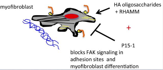
Fig1. HA oligosaccharides bind to RHAMM on the cell surface which regulates adhesion plaque signaling through FAK, promotes myoblast differentiation/migration. (Cornelia Tolg, 2012)
Not For Human Consumption!
Inquiry
- Reviews
- Q&As


Ask a Question for All HMMR Products
Required fields are marked with *
My Review for All HMMR Products
Required fields are marked with *
Inquiry Basket