Active Recombinant Human FGF3 Protein, Pre-aliquoted
| Cat.No. : | FGF3-03H |
| Product Overview : | Recombinant Human FGF3 Protein, Asp28 - Arg212, with an N-terminal Met, was expressed in E. coli. |
- Specification
- Gene Information
- Related Products
- Case Study
- Application
- Download
| Species : | Human |
| Source : | E.coli |
| Protein Length : | Asp28-Arg212, with an N-terminal Met |
| Description : | The protein encoded by this gene is a member of the fibroblast growth factor (FGF) family. FGF family members possess broad mitogenic and cell survival activities and are involved in a variety of biological processes including embryonic development, cell growth, morphogenesis, tissue repair, tumor growth and invasion. This gene was identified by its similarity with mouse fgf3/int-2, a proto-oncogene activated in virally induced mammary tumors in the mouse. Frequent amplification of this gene has been found in human tumors, which may be important for neoplastic transformation and tumor progression. Studies of the similar genes in mouse and chicken suggested the role in inner ear formation. |
| Bio-activity : | Measured in a cell proliferation assay using NR6R‑3T3 mouse fibroblast cells. The ED50 for this effect is 0.02-0.1 μg/mL in the presence of 1 μg/mL of heparin. |
| Molecular Mass : | Predicted Molecular Mass:21.1 kDa |
| N-terminal Sequence Analysis : | Met |
| Endotoxin : | < 0.01 EU/μg of the protein by the LAL method. |
| Purity : | > 97% by SDS-PAGE under reducing conditions and visualized by silver stain. |
| Stability : | Use a manual defrost freezer and avoid repeated freeze-thaw cycles. • 6 months from date of receipt at room temperature. • 12 months from date of receipt at 2-8 centigrade as supplied. • 1 month at 2-8 centigrade under sterile conditions after reconstitution. • 3 months at -20 to -80 centigrade under sterile conditions after reconstitution. |
| Reconstitution : | For a stock solution, reconstitute at 100 μg/mL in sterile PBS, or simply roll Pre-aliquoted directly to the medium for immediate use. |
| Shipping : | The product is shipped at ambient temperature. |
| Gene Name | FGF3 fibroblast growth factor 3 [ Homo sapiens (human) ] |
| Official Symbol | FGF3 |
| Synonyms | FGF3; fibroblast growth factor 3; INT2; HBGF-3; fibroblast growth factor 3; FGF-3; INT-2 proto-oncogene protein; V-INT2 murine mammary tumor virus integration site oncogene homolog; fibroblast growth factor 3 (murine mammary tumor virus integration site (v-int-2) oncogene homolog); heparin-binding growth factor 3; murine mammary tumor virus integration site 2, mouse; oncogene INT2; proto-oncogene Int-2 |
| Gene ID | 2248 |
| mRNA Refseq | NM_005247 |
| Protein Refseq | NP_005238 |
| MIM | 164950 |
| UniProt ID | P11487 |
| ◆ Cell & Tissue Lysates | ||
| FGF3-6240HCL | Recombinant Human FGF3 293 Cell Lysate | +Inquiry |
Case 1: Lamb R, et al. Oncotarget. 2015
Researchers created an isogenic cell model to uncover breast cancer biomarkers, using insights from the mouse mammary tumor virus (MMTV). By overexpressing WNT1 and FGF3 in MCF7 cells, they enhanced stem cell characteristics, including a significant increase in mammosphere formation. Proteomic analysis showed increased expression of EMT markers and proteins associated with mitochondrial activity, glycolysis, and protein synthesis, typical of anabolic cancer stem cells. MitoTracker staining validated the elevated mitochondrial mass induced by WNT1/FGF3. This model suggests that mitochondrial mass could serve as a biomarker for CSCs, shedding light on the importance of mitochondrial biogenesis in cancer cell propagation. This approach could be useful for biomarker discovery and targeting CSCs in various cancers.
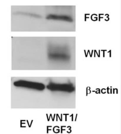
Fig1. Recombinant over-expression of WNT1 and FGF3 in these transfected cell models.
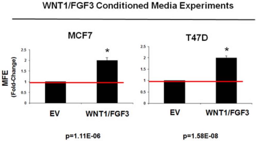
Fig2. Conditioned media from WNT1/FGF3 expressing MCF7 cells increases mammosphere formation.
Case 2: Sela-Donenfeld D, et al. BMC Dev Biol. 2009
In the development of the vertebrate CNS, the hindbrain forms rhombomeres, segmented units demarcated by boundary cells with unique traits like a stretched shape and lower proliferation. These cells' roles and formation controls are not well understood. This study found that boundary cells express signaling molecules like FGF3, which is normally transient in rhombomeres. Chick embryos with boundary cell deficiency due to truncated EphA4 receptor overexpression maintain FGF3 expression beyond its usual boundaries. Similarly, removing the r3/r4 boundary or blocking r4's contact with boundary cells led to persistent FGF3 expression, suggesting a role for boundary cells in segmental patterning.
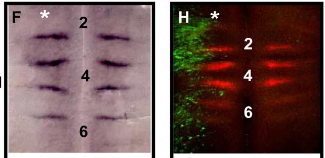
Fig1. FGF3 transcripts are restricted to boundaries in control electroporations.
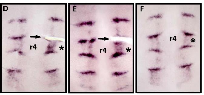
Fig2. Flat-mounted hindbrains of embryos in situ hybridized with FGF3 probe at 28 ss.
Recombinant Human FGF3 Protein, also known as fibroblast growth factor 3 (FGF-3), is a member of the fibroblast growth factor (FGF) family, which consists of at least 23 members. FGFs are heparin-binding growth factors that share a common tertiary structure due to a core 120 amino acid FGF domain. They play crucial roles in various physiological functions, including cell growth, angiogenesis, pattern formation, embryonic development, metabolic regulation, cell migration, neurotrophic effects, and tissue repair.
FGF-3, originally identified as a proto-oncogene, is involved in the development of the inner ear and is implicated in the synergistic regulation of dorsoventral axis formation during neuronal development. It binds with high affinity to the IIIb isoforms of FGF receptors 1 and 2 (FGFR1 and FGFR2), and also to the IIIc isoform of FGFR2, but with lower affinity.
In terms of practical applications, FGF-3 has been investigated for its potential roles in cancer development, particularly in bladder cancer. It is expressed in various malignancies and is considered a promising therapeutic target. Studies have shown that FGF-3 can induce cell proliferation and transformation, and its inhibition can lead to reduced tumor growth. Additionally, FGF-3 has been studied for its role in wound healing and tissue regeneration, with potential applications in the treatment of chronic wounds, ulcers, and burns.
The development of recombinant FGF-3 protein allows for detailed research into its functions and interactions with other components of the immune and nervous systems, which could lead to novel therapeutic strategies in various medical fields, including oncology and regenerative medicine.
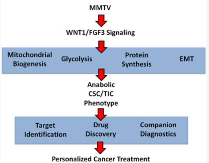
Fig1. Exploiting a humanized model of MMTV signaling to identify the characteristics of anabolic CSCs and achieve the goals of personalized medicine. (Rebecca Lamb, 2015)
Not For Human Consumption!
Inquiry
- Reviews
- Q&As


Ask a Question for All FGF3 Products
Required fields are marked with *
My Review for All FGF3 Products
Required fields are marked with *
Inquiry Basket