Active Recombinant Human CEACAM1 protein(Met1-Gly428), His&hFc-tagged
| Cat.No. : | CEACAM1-663H |
| Product Overview : | Recombinant Human CEACAM1 (NP_001020083.1) (Met 1-Gly 428) was expressed in HEK293, fused with the C-terminal polyhistidine-tagged Fc region of Human IgG1 at the C-terminus. |
| Availability | April 20, 2025 |
| Unit | |
| Price | |
| Qty |
- Specification
- Gene Information
- Related Products
- Case Study
- Application
- Download
| Species : | Human |
| Source : | HEK293 |
| Tag : | Fc&His |
| Protein Length : | Met1-Gly428 |
| Form : | Lyophilized from sterile 100 mM Glycine, 500 mM NaCl, 5 % Glycerol, 50 mM Arg Hcl, 50 mM Glu, 0.01 % tween 20, pH 5.0. Normally 5 % - 8 % trehalose, mannitol and 0.01% Tween80 are added as protectants before lyophilization. |
| Bio-activity : | Measured by its binding ability in a functional ELISA. Immobilized CEACAM8-His at 10 μg/mL (100 μL/well) can bind CEACAM1-Fch, the EC50 of CEACAM1-Fch is 70-200 ng/mL. |
| Molecular Mass : | The secreted recombinant human CEACAM1/Fc chimera is a disulfide-linked homodimer. The reduced monomer comprises 642 amino acids and predicts a molecular mass of 71.4 kDa. As a result of glycosylation, the apparent molecular mass of rhCEACAM1 is approximately 124.8 kDa in SDS-PAGE under reducing conditions. |
| Endotoxin : | < 1.0 EU per μg of the protein as determined by the LAL method. |
| Purity : | > 90 % as determined by SDS-PAGE |
| Storage : | Samples are stable for up to twelve months from date of receipt at -20°C to -80°C. Store it under sterile conditions at -20°C to -80°C. It is recommended that the protein be aliquoted for optimal storage. Avoid repeated freeze-thaw cycles. |
| Reconstitution : | It is recommended that sterile water be added to the vial to prepare a stock solution of 0.2 ug/ul. Centrifuge the vial at 4°C before opening to recover the entire contents. |
| Gene Name | CEACAM1 carcinoembryonic antigen-related cell adhesion molecule 1 (biliary glycoprotein) [ Homo sapiens ] |
| Official Symbol | CEACAM1 |
| Synonyms | CEACAM1; carcinoembryonic antigen-related cell adhesion molecule 1 (biliary glycoprotein); BGP; carcinoembryonic antigen-related cell adhesion molecule 1; BGP1; CD66a; antigen CD66; CD66a antigen; BGPI; |
| Gene ID | 634 |
| mRNA Refseq | NM_001024912 |
| Protein Refseq | NP_001020083 |
| MIM | 109770 |
| UniProt ID | P13688 |
| ◆ Recombinant Proteins | ||
| CEACAM1-6372Z | Recombinant Zebrafish CEACAM1 | +Inquiry |
| Ceacam1-881MAF647 | Recombinant Mouse Ceacam1 Protein, Fc-tagged, Alexa Fluor 647 conjugated | +Inquiry |
| CEACAM1-2650H | Recombinant Human CEACAM1 protein(151-230 aa), C-His-tagged | +Inquiry |
| CEACAM1-28H | Recombinant Human CEACAM1 protein, His-tagged | +Inquiry |
| CEACAM1-1091H | Recombinant Human CEACAM1 Protein, GST-Tagged | +Inquiry |
| ◆ Cell & Tissue Lysates | ||
| CEACAM1-3039HCL | Recombinant Human CEACAM1 cell lysate | +Inquiry |
| CEACAM1-2214MCL | Recombinant Mouse CEACAM1 cell lysate | +Inquiry |
| CEACAM1-1239RCL | Recombinant Rat CEACAM1 cell lysate | +Inquiry |
Case 1: Zhu Y, et al. J Transl Med. 2019
Interferon gamma (IFNγ) is a key player in chronic lung diseases, primarily by boosting inflammation through increased interleukin production. Our study looked into how this happens, focusing on the interaction between CEACAM1 and the PI3K pathway in lung cells. When IFNγ was introduced, it ramped up the levels of interleukins IL6 and IL8 and enhanced CEACAM1 expression, particularly the CEACAM1-S isoforms, in bronchial cells. This not only spurred cell growth and movement but also set off a feedback loop where CEACAM1 and the PI3K/Akt/mTOR pathway activate each other, driving further inflammatory responses.
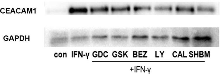
Fig1. Protein levels of CEACAM1 in HBE cells.
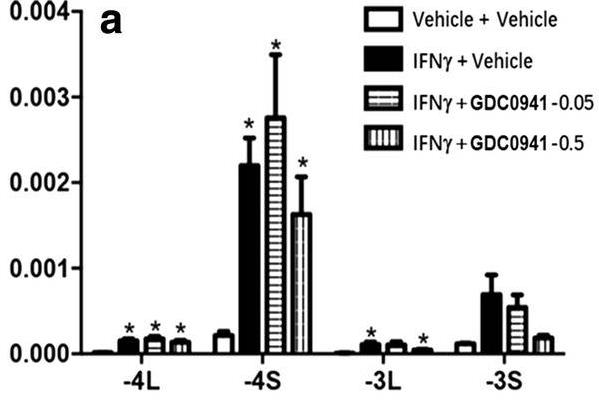
Fig2. Effect of PI3K/Akt pathway inhibitors on IFN-γ induced CEACAM1 and its isoform expression.
Case 2: Xu J, et al. Cell Physiol Biochem. 2018
CEACAM1, a molecule from the CEA family known as CD66a, has a complex influence on cancer behavior. In the context of multiple myeloma, ramping up CEACAM1 levels in specific cell lines led to decreased cell growth, more apoptosis, and reduced ability of cells to invade and move. This effect seems tied to activating caspase-3 and reducing MMP-2 and MMP-9. Notably, CEACAM1 appeared more frequently in patients with early-stage disease and those with lower levels of β2-microglobulin.
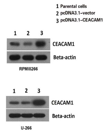
Fig1. CEACAM1 overexpression in U-266 and RPMI8266 cell lines was confirmed at the protein level by Western blot.
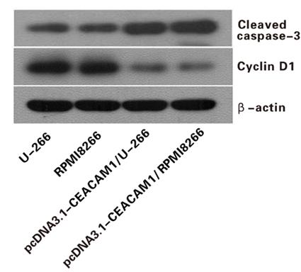
Fig2. western blotting analysis showed that caspase-3 was activated but cyclinD1 was inhibited after CEACAM1 transfection in both U-266 and RPMI8266 cell lines.
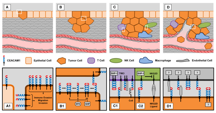
Fig1. CEACAM1 in different stages of tumorigenesis and progression. (Lisa Götz, 2023)
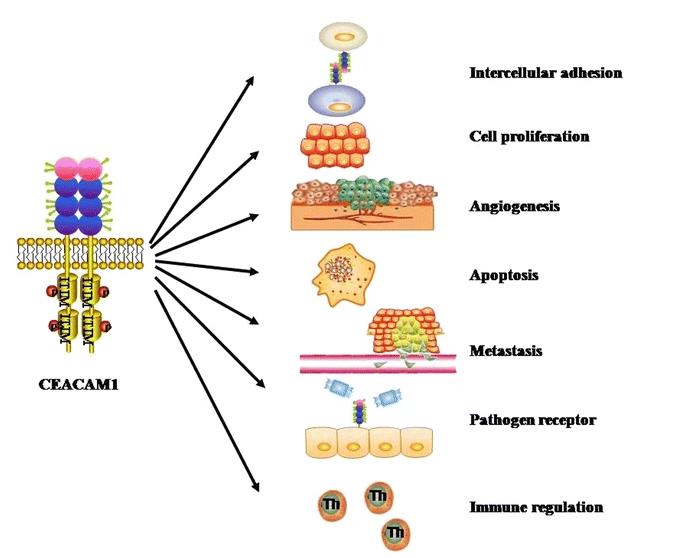
Fig2. The functions of CEACAM1. (Ye Ling, 2015)
Not For Human Consumption!
Inquiry
- Reviews
- Q&As


Ask a Question for All CEACAM1 Products
Required fields are marked with *
My Review for All CEACAM1 Products
Required fields are marked with *
Inquiry Basket