Purified fluorescent protein ECFP
| Cat.No. : | ECFP-20 |
| Product Overview : | Purified fluorescent protein ECFP, with N-terminal HIS tag, expressed in E. coli, 250ug |
- Specification
- Gene Information
- Related Products
- Case Study
- Application
- Download
| Source : | E.coli |
| Tag : | His |
| Molecular Mass : | 26.9 kDa |
| AA Sequence : | MVSKGEELFTGVVPILVELDGDVNGHKFSVSGEGEGDATYGKLTLKFICTTGKLPVPWPTLVTTLTWGVQCFSRY PDHMKQHDFFKSAMPEGYVQERTIFFKDDGNYKTRAEVKFEGDTLVNRIELKGIDFKEDGNILGHKLEYNYISHN VYITADKQKNGIKANFKIRHNIEDGSVQLADHYQQNTPIGDGPVLLPDNHYLSTQSALSKDPNEKRDHMVLLEFV TAAGITLGMDELYK |
| Purity : | >80% as determined by SDS-PAGE and Coomassie blue staining |
| Concentration : | >50 ug/mL as determined by microplate BCA method |
| Storage Buffer : | PBS, pH7.4, 10% glycerol. |
Case 1: Xie C, et al. J Orthop Res. 2024
In orthopedics, the "Race for the Surface" theory, which suggests a competition between bacteria and host cells on an implant post-surgery, lacks live in vivo confirmation. To address this, researchers enhance d a mouse imaging system to track green fluorescent protein (GFP+) host cells and enhanced cyan fluorescent protein (ECFP+) or red fluorescent protein (RFP+) MRSA near a femoral implant. After inoculation with ~10^5 CFU, an L-shaped implant was fitted beneath a GRIN lens. Initial imaging showed scant bacterial presence, but by 3 hours, ECFP+ MRSA covered 56.8% of the implant surface. 3D imaging confirmed exponential MRSA growth from 3 to 6 hours, corroborated by a significant increase in bacterial load on the implant from 2 to 12 hours post-operation.
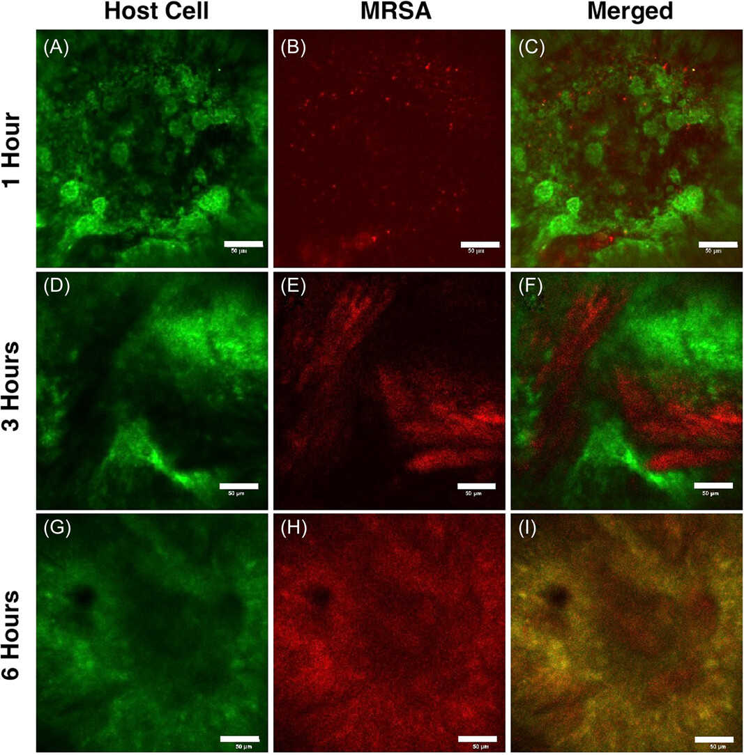
Fig1. Representative longitudinal intravital imaging of the bone marrow (LIMB) of the Race for the Surface.
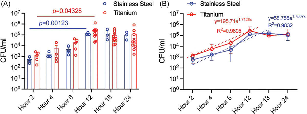
Fig2. Ex vivo assessment of methicillin-resistant Staphylococcus aureus (MRSA) bacterial load and growth on the implant over 24 h.
Case 2: Amreen, et al. Biology (Basel). 2020
Hydrogen peroxide (H2O2) plays a key role in cell signaling and metabolism, and its levels rise under stress, leading to oxidative stress in plants and animals. To study H2O2 dynamics, researchers created the FLIP-H2O2 nanosensor using a FRET-based approach with ECFP and mVenus flanking the OxyR regulatory domain. This pH-stable sensor is highly selective for H2O2 and insensitive to other oxidants. It has a broad dynamic range and a Kd of 247 μM, localizing to the cytosol in various cell types. The FLIP-H2O2 allows for non-invasive, real-time monitoring of H2O2 levels through FRET-ratio changes in response to stress or added ligand.
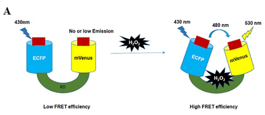
Fig1. Regulatory domain is attached with donor (ECFP) and acceptor (mVenus).
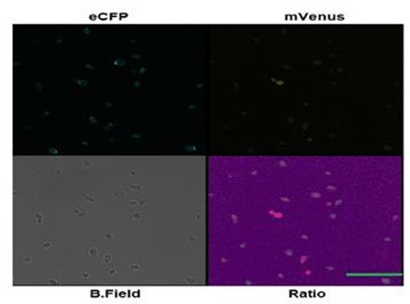
Fig2. Confocal microscopy imaging of nanosensor expressing yeast cells showing ECFP, mVenus, bright field and ratio images at various channels.
ECFP (Enhanced Green Fluorescent Protein) is a fluorescent protein, the brightness and light stability of ECFP is a key indicator of its application value, and it is often used as a marker in vivo to observe and track the activity of cells or proteins. In addition, it has other extensive applications in bioimaging and molecular biology research.
Fluorescence Resonance Energy Transfer (FRET) : ECFP is a common donor in FRET experiments, allowing the study of protein-protein interactions and other biological events by monitoring the efficiency of energy transfer between the donor and recipient. FRET technology is widely implemented in living cells to study protein-protein interactions and to monitor intracellular processes such as calcium wave induction, pH changes, and so on through biosensors.
Biosensors: ECFP is used to develop a variety of biosensors that can report important processes within cells such as calcium concentration changes, pH changes, membrane potential fluctuations, and more. For example, by fusing pairs of fluorescent proteins onto biopolymers, molecular probes for optically living cell imaging can be developed.
Intermolecular measurements: In addition to applications in biosensors, ECFP can also be used for intermolecular measurements, for example, by fusing fluorescent proteins onto two separate proteins, the interaction between them can be studied.
Optically illuminated fluorescent protein: In live cell imaging and protein dynamics studies, ECFP can be used as an optically illuminated fluorescent protein to produce a higher level of contrast through light activation or light transformation, suitable for photobleaching techniques.
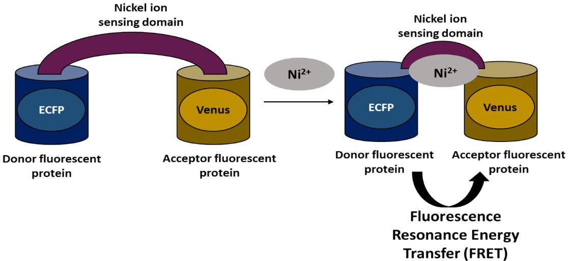
Fig1. Schematic representation of FRET. (Neha Soleja, 2019)
Not For Human Consumption!
Inquiry
- Reviews
- Q&As


Ask a Question for All ECFP Products
Required fields are marked with *
My Review for All ECFP Products
Required fields are marked with *
Inquiry Basket