Butyrophilins
Related Symbol Search List
Immunology Background
Background
About Butyrophilins
Butyrophilins (BTNs) are a family of transmembrane glycoproteins that belong to the immunoglobulin superfamily. BTNs are type I transmembrane proteins with a similar overall structure. They consist of an extracellular region that contains variable numbers of Ig-like domains, a transmembrane domain, and a cytoplasmic domain that often contains a B30.2 domain. The extracellular Ig-like domains are involved in protein-protein interactions, while the B30.2 domain is thought to have diverse functions, including possible roles in ligand binding and immune modulation.
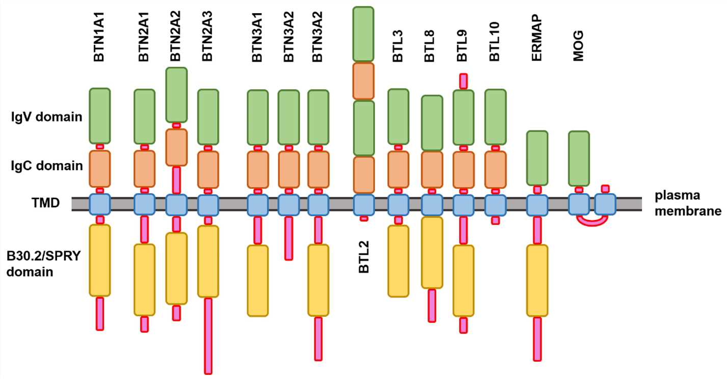 Fig.1 Schematic representation of the modular domain structure of the members of human butyrophilin family. (Redwan EM, et al., 2018)
Functional Characteristics
BTNs play important roles in immune regulation and immune cell interactions. Their functions include:
- Modulation of T cell responses: BTNs can interact with T cells and modulate their activation, differentiation, and effector functions. They can act as co-stimulatory or co-inhibitory molecules, depending on the specific context and interaction partners.
- Immune tolerance and regulation: BTNs are involved in maintaining immune tolerance and preventing excessive immune responses. They contribute to the balance between immune activation and immune regulation, ensuring proper immune function.
- Interactions with other immune molecules: BTNs can interact with various immune molecules, including TCRs, co-stimulatory receptors, and co-inhibitory receptors. These interactions influence immune cell signaling and the outcome of immune responses.
- Involvement in diseases: Dysregulation of BTNs has been associated with various diseases, including autoimmune disorders, infectious diseases, and cancer. Understanding the roles of BTNs in disease pathogenesis may lead to the development of novel therapeutic approaches.
Co-stimulatory and Co-inhibitory Pathways of the Butyrophilins
The co-stimulatory and co-inhibitory pathways of the BTNs are still an area of active research, and their roles in immune regulation are not yet fully understood. However, emerging evidence suggests that certain BTNs can function as co-stimulatory or co-inhibitory molecules in immune responses. Here is an overview of the co-stimulatory and co-inhibitory pathways associated with BTNs:
| Pathways | BTNs and functions | |
|---|---|---|
| Co-stimulatory Pathways | BTNL2 - Unknown Receptors |
|
| Co-inhibitory Pathways | BTN3A1 (CD277) - Vγ9Vδ2 TCR |
|
| BTN3A1 - Butyrophilin-Associated Molecules (BAMs) |
|
|
It's important to note that the specific mechanisms and signaling pathways associated with BTNs and their interactions are still being actively investigated. The functions of BTNs in co-stimulation and co-inhibition are likely to be context-dependent and may vary depending on the specific BTN isoforms, interacting receptors, and cell types involved.
Further research is needed to elucidate the precise roles of BTNs in co-stimulatory and co-inhibitory pathways and their implications for immune regulation and disease. However, the emerging understanding of BTNs' involvement in immune responses highlights their potential as targets for therapeutic interventions aimed at modulating immune activation or tolerance.
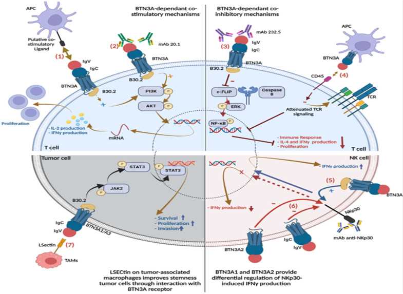 Fig.2 BTN3A interaction patterns and signaling pathways. BTN3A-dependent co-stimulatory mechanisms. (Kone AS, et al., 2022)
BTN Famliy Molecules
BTNs can be classified into three main subfamilies:
| Key molecule types | Functions |
|---|---|
| Butyrophilin proteins |
|
| Butyrophilin-like proteins (BTNL) |
|
| SKINT-like factor |
|
These additional groups of molecules expand the diversity of the butyrophilin family. They contribute to immune regulation, T cell responses, and antigen recognition in different contexts. Further research is needed to fully understand their functions and roles in immunity.
Role of Butyrophilins in Different Diseases
BTNs have been implicated in various diseases, reflecting their diverse roles in immune regulation and other biological processes. Here are some examples of the involvement of BTNs in different diseases:
Autoimmune Diseases
- Several BTNs, particularly the B7-CD28-related butyrophilins (BTNLs), have been associated with autoimmune diseases such as rheumatoid arthritis, multiple sclerosis, and inflammatory bowel disease.
- Dysregulation of BTNL expression or function can affect the balance between immune activation and regulation, contributing to autoimmune pathology.
Infectious Diseases
- BTNs, such as BTNL2 and BTN3A1, have been implicated in infectious diseases.
- BTNL2 has been linked to chronic hepatitis B virus infection and has been proposed as a potential biomarker for disease progression.
- BTN3A1, specifically its interaction with the Vγ9Vδ2 T cell receptor (TCR), plays a role in the recognition of microbial metabolites produced by certain bacteria and parasites. This interaction can influence the immune response against infections.
Cancer
- BTNs have been associated with various aspects of cancer, including tumor development, progression, and immune evasion.
- Some BTNs, such as BTN1A1, have been implicated in breast cancer. Alterations in BTN1A1 expression have been observed in breast cancer tissues and are associated with poor prognosis.
- BTNL1 and BTNL6 have been found to be upregulated in certain types of cancer, including lung cancer and melanoma, and their overexpression has been correlated with tumor progression and reduced patient survival.
Inflammatory Disorders
- BTNs, particularly BTNLs, have been linked to inflammatory disorders such as inflammatory bowel disease (IBD) and psoriasis.
- Genetic variations in BTNL2 have been associated with an increased risk of developing IBD.
- BTNL2 has also been implicated in the pathogenesis of psoriasis, a chronic skin inflammatory disorder.
It's important to note that the specific roles of BTNs in disease pathogenesis are still being actively investigated, and our understanding of their contributions to different diseases is evolving. The complex interactions between BTNs, immune cells, and disease processes require further research to unravel the underlying mechanisms.
Studying the involvement of BTNs in various diseases may provide insights into disease mechanisms, identify potential diagnostic markers, and uncover therapeutic targets for intervention. However, more research is needed to fully elucidate the precise roles of BTNs in specific disease contexts and to explore their therapeutic potential.
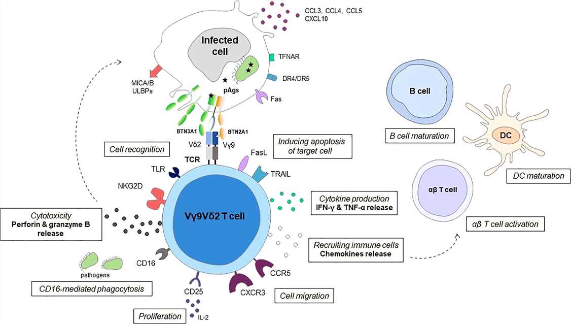 Fig.3 Schematic representation of effector mechanisms of Vγ9Vδ2 T cells in response to infection. (Gay L, et al., 2022)
Case Study
Case 1: Vantourout P, Laing A, Woodward M J, et al. Heteromeric interactions regulate butyrophilin (BTN) and BTN-like molecules governing γδ T cell biology. Proceedings of the National Academy of Sciences, 2018, 115(5): 1039-1044.
The authors assessed whether the intracellular structural domains of BTN3A1 and BTN3A2 contribute to ER retention/retrieval. FLAG-tagged BTN3A1 introduced into CRA123 cells largely colocalized with the ER marker, protein disulphide isomerase (PDI), but not with a Golgi marker, Gm130 (A), quantitated by high bright detail similarity (BDS) of FLAG-PDI, but not of FLAG-Gm130 (A). Just as BTN3A2 enhanced BTN3A1 surface expression, BTN3A1 showed reduced ER colocalization in cells cotransfected with BTN3A2 (Fig. 5A, orange segregation from cyan; reduced FLAG-PDI BDS). Confocal microscopy likewise revealed that mCherry-tagged Sec61β, another ER-resident marker, colocalized with cotransfected BTN3A1 or BTN3A2 expressed singly, but less so when BTN3A1 and BTN3A2 were coexpressed (B).
 Fig.1 BTN3A1 and BTN3A2 associate with the ER and regulate each other's trafficking.
Case 2: Kim YS, Lee SH, Park AH, et al. BTN1A1 is a novel immune checkpoint mutually exclusive to PD-L1. J Immunother Cancer. 2024;12(3):e008303.
BTN1A1 can expressed on tumor cells, and function to suppress T-cell responses in vitro and in vivo. The author thus sought to conduct a more in-depth analysis of the role of tumor-derived BTN1A1 as a novel immune checkpoint. To gauge the potential feasibility of BTN1A1 blockade, the author developed a rabbit monoclonal antibody (STC43H11-1) for IHC staining (A, B) and used it to visualize BTN1A1 expression in a TMA. Pronounced and robust BTN1A1 expression was evident in head and neck, bladder, squamous non-small cell lung cancer (NSCLC), ovarian, cervical, prostate, and other tumor tissues, while PD-L1 expression was relatively low in both frequency and intensity (C, D). Simultaneous PD-L1 and BTN1A1 detection using an OPAL multiplex system revealed that while these proteins were sometimes present on adjacent cells, they were exclusively expressed on a given cell in head, neck, colon, and ovarian tumors (E-G). The author additionally quantified the number of BTN1A1+PD-L1−, BTN1A1− PD-L1+, BTN1A1− PD-L1−, or BTN1A1+PD-L1+ cells within each patient specimen as a percentage of cancer cells expressing cytokeratin (H), supporting this observation.
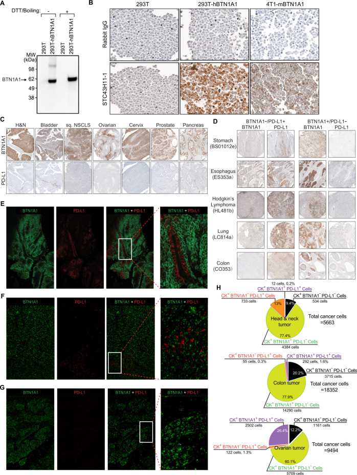 Fig.2 BTN1A and PD-L1 exhibit mutually exclusive expression in tumor cells.
References
- Redwan EM, Al-Hejin AM, Almehdar HA, Elsaway AM, Uversky VN. Prediction of disordered regions and their roles in the anti-pathogenic and immunomodulatory functions of butyrophilins. Molecules. 2018; 23(2):328.
- Rigau M, Uldrich AP, Behren A. Targeting butyrophilins for cancer immunotherapy. Trends Immunol. 2021;42(8):670-680.
- Kone AS, Ait Ssi S, Sahraoui S, Badou A. BTN3A: A promising immune checkpoint for cancer prognosis and treatment. Int J Mol Sci. 2022;23(21):13424.
- Gay L, Mezouar S, Cano C, et al. Role of Vγ9vδ2 T lymphocytes in infectious diseases. Front Immunol. 2022;13:928441.