
OPA1
-
Official Full Name
optic atrophy 1 (autosomal dominant) -
Overview
This gene product is a nuclear-encoded mitochondrial protein with similarity to dynamin-related GTPases. It is a component of the mitochondrial network. Mutations in this gene have been associated with optic atrophy type 1, which is a dominantly inherited optic neuropathy resulting in progressive loss of visual acuity, leading in many cases to legal blindness. Multiple transcript variants encoding different isoforms have been found for this gene. -
Synonyms
OPA1;optic atrophy 1 (autosomal dominant);dynamin-like 120 kDa protein, mitochondrial;dynamin like guanosine triphosphatase;FLJ12460;KIAA0567;MGM1;mitochondrial dynamin like GTPase;NPG;NTG;optic atrophy protein 1;mitochondrial dynamin-like GTPa
Recombinant Proteins
- Zebrafish
- Human
- Chicken
- Rat
- Mouse
- Pongo Abelii
- Oncorhynchus masou (Cherry salmon) (Masu salmon)
- zebrafish
- Mammalian Cells
- Wheat Germ
- HEK293
- E.coli
- His
- Non
- Avi
- Fc
- Flag
- DDK
- Myc
Background
What is OPA1 protein?
OPA1 gene (OPA1 mitochondrial dynamin like GTPase) is a protein coding gene which situated on the long arm of chromosome 3 at locus 3q29. The OPA1 protein (Optic Atrophy 1) is a protein that is active in the mitochondria of cells. Mitochondria are the energy-producing centers of the cell and are dynamic structures that change shape through the processes of division (fission) and fusion (fusion), which are essential for mitochondrial function and the production of new mitochondria. The OPA1 protein helps regulate the shape of mitochondria by playing a key role in the process of mitochondrial fusion. In addition, OPA1 proteins are involved in the oxidative phosphorylation process in mitochondria, which is an important way for cells to obtain energy. The OPA1 protein is also involved in maintaining DNA within mitochondria (called mitochondrial DNA or mtDNA) and plays a role in regulating cell death (apoptosis). The OPA1 protein is consisted of 960 amino acids and OPA1 molecular weight is approximately 111.6 kDa.
What is the function of OPA1 protein?
OPA1 protein is an important factor that promotes inner mitochondrial fusion and helps to form mitochondrial network. OPA1 regulates mitochondrial morphology by participating in the process of mitochondrial division and fusion, which is essential for mitochondrial function. OPA1 is involved in the process of oxidative phosphorylation in mitochondria, which is the primary pathway by which cells produce energy. OPA1 proteins are also involved in the maintenance of DNA within mitochondria, which is important for mitochondrial function and cellular respiration. OPA1 also plays a role in the process of mitochondrial mediated programmed cell death (apoptosis). OPA1 indirectly affects the metabolic state of cells, including metabolism and polarization in macrophages, by maintaining mitochondrial morphology and function.
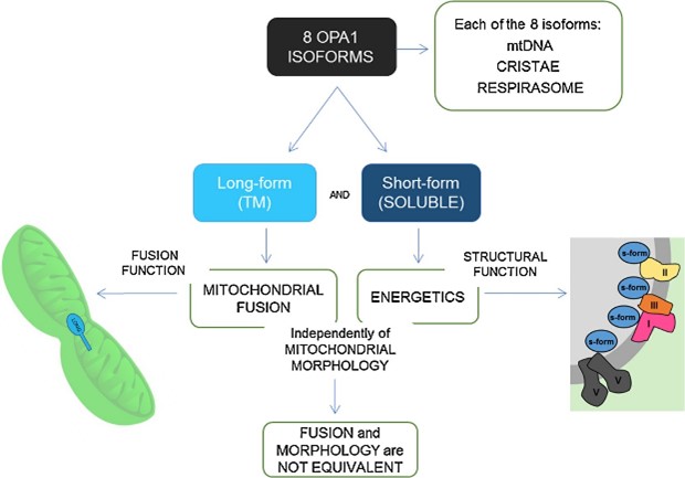
Fig1. Summary of OPA1 isoforms functions. (Valentina Del Dotto, 2018)
OPA1 Related Signaling Pathway
OPA1 is a key regulator of mitochondrial fusion and interacts with mitochondrial division factors such as Drp1 (Dynamin-related protein 1) to maintain the dynamic balance of mitochondrial morphology and function. The AKT-GSK3β signaling pathway influences mitochondrial function and apoptosis by regulating OPA1 clipping, especially in oxidative stress-induced apoptosis. OPA1 affects mitochondrial metabolism and cell polarization by influencing mitochondrial morphology, especially in immune cells such as macrophages. OPA1 indirectly influences the metabolic state of cells by maintaining mitochondrial morphology and function, including metabolic pathways in specific cell types, such as in skeletal muscle and cardiac muscle.
OPA1 Related Diseases
OPA1 diseases are diverse, especially those closely linked to mitochondrial morphology and function. OPA1 gene mutation is the Dominant locus of Dominant Optic Atrophy (DOA). The condition is characterized by progressive vision loss, usually beginning in childhood or adolescence, and may be accompanied by hearing impairment. Abnormalities in OPA1 have also been linked to other neurodegenerative diseases, such as Parkinson's disease. In addition, mutations in the OPA1 gene may affect the auditory nerve, leading to auditory neuropathy. OPA1 plays an important role in the process of apoptosis, and its abnormality may be related to the occurrence and development of some cancers. OPA1 is also involved in heat shock response and may play a role in toxic inhibition of anticancer drugs. In addition, OPA1 is associated with mitochondrial dysfunction and may play a role in smoking-induced chronic obstructive pulmonary disease (COPD).
Bioapplications of OPA1
OPA1 mutations are associated with specific genetic disorders such as optic atrophy (DOA), so genetic testing for OPA1 can be used as a tool to diagnose these disorders. OPA1 is a potential therapeutic target for diseases associated with mitochondrial dysfunction, and drug development for OPA1 may help treat diseases associated with mitochondrial dysfunction, such as certain types of neurodegenerative diseases. Gene therapy, functional studies of the OPA1 gene offer the possibility of developing gene therapies targeting genetic diseases caused by OPA1 mutations.
Case Study
Case Study 1: Chun-Xia Yu, 2024
Cisplatin resistance is a major obstacle in the treatment of non-small cell lung cancer (NSCLC). p32 and OPA1 are the key regulators of mitochondrial morphology and function. This study aims to investigate the role of the p32/OPA1 axis in cisplatin resistance in NSCLC and its underlying mechanism. The levels of p32 protein and mitochondrial fusion protein OPA1 are higher in cisplatin-resistant A549/DDP cells than in cisplatin-sensitive A549 cells, which facilitates mitochondrial fusion in A549/DDP cells. In addition, the expression of p32 and OPA1 protein is also upregulated in A549 cells during the development of cisplatin resistance. Furthermore, metformin significantly downregulates the expressions of p32 and OPA1 and induces mitochondrial fission and a decrease in ATP level in A549/DDP cells. Moreover, D-erythro-Sphingosine, a potent p32 kinase activator, counteracts the metformin-induced downregulation of OPA1 and mitochondrial fission in A549/DDP cells.
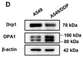
Fig1. Expressions of Drp1 and OPA1 in A549 and A549/DDP cells.
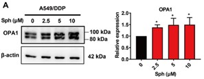
Fig2. Effect of D-erythro-Sphingosine on metformin-induced downregulation of OPA1 in A549/DDP cells.
Case Study 2: Alexander von der Malsburg, 2023
Distinct morphologies of the mitochondrial network support divergent metabolic and regulatory processes that determine cell function and fate1-3. The mechanochemical GTPase optic atrophy 1 (OPA1) influences the architecture of cristae and catalyses the fusion of the mitochondrial inner membrane4,5. Despite its fundamental importance, the molecular mechanisms by which OPA1 modulates mitochondrial morphology are unclear. Here, using a combination of cellular and structural analyses, researchers illuminate the molecular mechanisms that are key to OPA1-dependent membrane remodelling and fusion. Human OPA1 embeds itself into cardiolipin-containing membranes through a lipid-binding paddle domain. A conserved loop within the paddle domain inserts deeply into the bilayer, further stabilizing the interactions with cardiolipin-enriched membranes. OPA1 dimerization through the paddle domain promotes the helical assembly of a flexible OPA1 lattice on the membrane, which drives mitochondrial fusion in cells. Moreover, the membrane-bending OPA1 oligomer undergoes conformational changes that pull the membrane-inserting loop out of the outer leaflet and contribute to the mechanics of membrane remodelling.
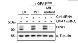
Fig3. The expression and stability of the siRNA-resistant OPA1 proteins were assessed.
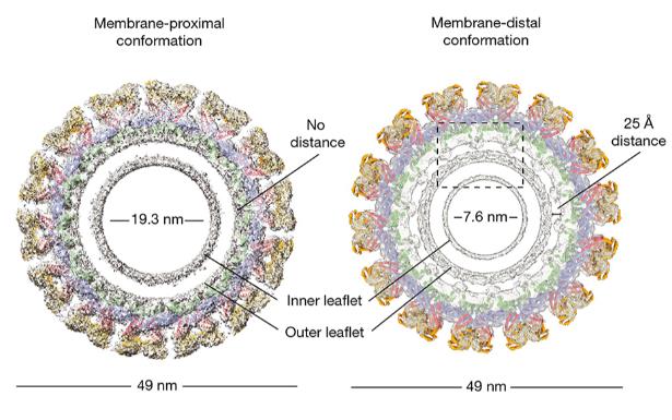
Fig4. Structural comparison of the membrane-proximal and membrane-distal conformations of the S-OPA1 polymer.
Quality Guarantee
High Purity
.jpg)
Fig1. SDS-PAGE (OPA1-3996H)
.
.jpg)
Fig2. SDS-PAGE (OPA1-4769H)
Involved Pathway
OPA1 involved in several pathways and played different roles in them. We selected most pathways OPA1 participated on our site, such as , which may be useful for your reference. Also, other proteins which involved in the same pathway with OPA1 were listed below. Creative BioMart supplied nearly all the proteins listed, you can search them on our site.
| Pathway Name | Pathway Related Protein |
|---|
Protein Function
OPA1 has several biochemical functions, for example, GTP binding,GTPase activity,magnesium ion binding. Some of the functions are cooperated with other proteins, some of the functions could acted by OPA1 itself. We selected most functions OPA1 had, and list some proteins which have the same functions with OPA1. You can find most of the proteins on our site.
| Function | Related Protein |
|---|---|
| magnesium ion binding | ATP10A,PKMA,FDXACB1,DUT,PRKACB,EPHX2,STK3,STK4,RPS6KA4,HACL1 |
| GTPase activity | GNAO1,RAB11A,GPN1,RAB7A,GNAV1,MRAS,GBP3,RAP2A,RAB3AA,DNM1A |
| protein binding | ARHGEF6,BOP1,PINK1,NELL2,PRX,TNK2,CRIPT,CD86,PI4KA,ZNF397 |
| GTP binding | RAP1B,RAB6A,GTPBP5,RAB11A,GPN3,CDC42L,ACSM1,RALBB,RAB3DB,GLUD1 |
Interacting Protein
OPA1 has direct interactions with proteins and molecules. Those interactions were detected by several methods such as yeast two hybrid, co-IP, pull-down and so on. We selected proteins and molecules interacted with OPA1 here. Most of them are supplied by our site. Hope this information will be useful for your research of OPA1.
BNIP3;MYC;SIRT3;LRRK2;KAT2B;PTP4A3
Resources
Related Services
Related Products
References
- Degardin, A; Dobbelaere, D; et al. Spinocerebellar Ataxia: A Rational Approach to Aetiological Diagnosis. CEREBELLUM 11:289-299(2012).
- Hanein, S; Garcia, M; et al. TMEM126A is a mitochondrial located mRNA (MLR) protein of the mitochondrial inner membrane. BIOCHIMICA ET BIOPHYSICA ACTA-GENERAL SUBJECTS 1830:3719-3733(2013).

