SYN1
-
Official Full Name
synapsin I -
Overview
This gene is a member of the synapsin gene family. Synapsins encode neuronal phosphoproteins which associate with the cytoplasmic surface of synaptic vesicles. Family members are characterized by common protein domains, and they are implicated in synaptogenesis and the modulation of neurotransmitter release, suggesting a potential role in several neuropsychiatric diseases. This member of the synapsin family plays a role in regulation of axonogenesis and synaptogenesis. The protein encoded serves as a substrate for several different protein kinases and phosphorylation may function in the regulation of this protein in the nerve terminal. Mutations in this gene may be associated with X-linked disorders with primary neuronal degeneration such as Rett syndrome. Alternatively spliced transcript variants encoding different isoforms have been identified. [provided by RefSeq, Jul 2008] -
Synonyms
SYN1;synapsin I;SYNI;SYN1a;SYN1b;synapsin-1;brain protein 4.1
Recombinant Proteins
- Human
- Rat
- Zebrafish
- Mouse
- E.coli
- HEK293
- Mammalian Cell
- Yeast
- Mammalian cells
- HEK293T
- E. coli
- Mamanlian cells
- GST
- Myc&DDK
- His
- His&T7
- His&Fc&Avi
- Flag
Background

Fig1. Phosphorylation sites of synapsin I by various protein kinases. (Yoko Yamagata, 2003)
What is SYN1 protein?
SYN1 (synapsin I) gene is a protein coding gene which situated on the short arm of chromosome X at locus Xp11. This gene is a member of the synapsin gene family. Synapsins encode neuronal phosphoproteins which associate with the cytoplasmic surface of synaptic vesicles. Family members are characterized by common protein domains, and they are implicated in synaptogenesis and the modulation of neurotransmitter release, suggesting a potential role in several neuropsychiatric diseases. This member of the synapsin family plays a role in regulation of axonogenesis and synaptogenesis. The protein encoded serves as a substrate for several different protein kinases and phosphorylation may function in the regulation of this protein in the nerve terminal. The SYN1 protein is consisted of 705 amino acids and its molecular mass is approximately 74.1 kDa.
What is the function of SYN1 protein?
The SYN1 protein, also known as synapsin I, is a neuronal phosphoprotein that is associated with the membranes of small synaptic vesicles. SYN1 is involved in the process of synaptic transmission, which is the communication between neurons through the release of neurotransmitters from synaptic vesicles at the synapse. It contributes to the development of neurons, particularly in the growth of neurites and the formation of synapses, which are essential for establishing neural networks in the brain. SYN1 is implicated in the creation of synapses, which are the junctions through which neurons signal to each other. The protein is also involved in maintaining the structure and function of mature synapses, which is important for the long-term stability of neural circuits. SYN1 plays a role in synaptic plasticity, which is the ability of synapses to strengthen or weaken over time, thus enabling learning and memory.
SYN1 Related Signaling Pathway
SYN1, or synapsin I, is a protein that plays a critical role in the regulation of synaptic vesicle trafficking and neurotransmitter release at nerve terminals. It is involved in several signaling pathways that are essential for normal neuronal function. Synapsins are implicated in signal transduction pathways that lead to the activation of various intracellular cascades upon synaptic stimulation. SYN1 overexpression has been shown to activate the cAMP signaling pathway, leading to the phosphorylation of CREB and PKA proteins, which in turn promote the secretion of neurotransmitters such as acetylcholine (ACh), dopamine (DA), and serotonin (5-HT). SYN1 is important for neuronal development and axonogenesis, as indicated by studies showing that synapsin I deficiency in mice leads to severe retardation in the outgrowth of axons and a delay in synapse formation.
SYN1 Related Diseases
Variants in the SYN1 gene are linked to a range of neurodevelopmental disorders that predominantly affect males. These may include conditions like intellectual disability and autism spectrum disorder. SYN1 has been implicated in the pathogenesis of certain forms of epilepsy. For instance, novel SYN1 variants have been reported in individuals with focal epilepsy. Mutations in the SYN1 gene may be associated with X-linked dominant primary neuronal degeneration diseases such as Rett syndrome, which is characterized by severe cognitive and motor impairments. The SYN1 gene is mentioned in the context of autism spectrum disorder, which is a developmental disorder affecting communication and behavior. SYN1's involvement in neuronal signaling could theoretically link it to conditions involving abnormal pain signaling, although this would require further substantiation.
Bioapplications of SYN1
SYN1 could potentially be a therapeutic target for the development of drugs aimed at modulating synaptic transmission in various neurological conditions. ariants or altered levels of SYN1 may serve as biomarkers for specific neurological conditions, aiding in diagnosis and disease progression monitoring. Understanding the function of SYN1 could inform the development of gene therapy approaches for disorders where SYN1 function is impaired. SYN1's involvement in synaptic vesicle trafficking could make it a target for pharmacological research into drugs that affect neurotransmission.
Case Study
Case Study 1: Jacques Gonzales, 2020
Most of the gut functions are controlled by the enteric nervous system (ENS), a complex network of enteric neurons located throughout the wall of the gastrointestinal tract. The formation of ENS connectivity during the perinatal period critically underlies the establishment of gastrointestinal motility, but the factors involved in this maturation process remain poorly characterized. Here, the researchers examined the role of Semaphorin 3A (Sema3A) on ENS maturation and its potential implication in Hirschsprung disease (HSCR), a developmental disorder of the ENS with impaired colonic motility. They found that Sema3A and its receptor Neuropilin 1 (NRP1) are expressed in the rat gut during the early postnatal period. At the cellular level, NRP1 is expressed by enteric neurons, where it is particularly enriched at growth areas of developing axons. Treatment of primary ENS cultures and gut explants with Sema3A restricts axon elongation and synapse formation. Comparison of the ganglionic colon of HSCR patients to the colon of patients with anorectal malformation shows reduced expression of the synaptic molecule synapsin 1 in HSCR, which is inversely correlated with Sema3A expression.
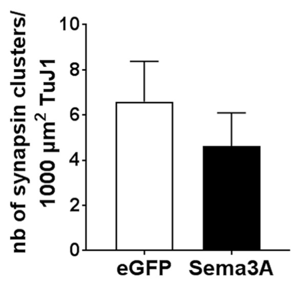
Fig1. Quantification of the number of synapsin 1 clusters.

Fig2. Linear regression analysis between Sema3A and synapsin 1 expression in HSCR colon.
Case Study 2: Anna Rocchi, 2019
Synapsin I is a phosphoprotein that coats the cytoplasmic side of synaptic vesicles and regulates their trafficking within nerve terminals. Autoantibodies against Syn I have been described in sera and cerebrospinal fluids of patients with numerous neurological diseases, including limbic encephalitis and clinically isolated syndrome; however, the effects and fate of autoantibodies in neurons are still unexplored. The researchers found that in vitro exposure of primary hippocampal neurons to patient's autoantibodies to SynI decreased the density of excitatory and inhibitory synapses and impaired both glutamatergic and GABAergic synaptic transmission. These effects were reproduced with a purified SynI antibody and completely absent in SynI knockout neurons. Autoantibodies to SynI are internalized by FcγII/III-mediated endocytosis, interact with endogenous SynI, and promote its sequestration and intracellular aggregation. Neurons exposed to human autoantibodies to SynI display a reduced density of SVs, mimicking the SynI loss-of-function phenotype.

Fig3. Quantification of the density of SynI immunoreactive synaptic puncta.

Fig4. Quantification of the immunoreactivity intensity of internalized SynI-mAb.
Quality Guarantee
High Purity
.jpg)
Fig1. SDS-PAGE (SYN1-1380H)
.
.jpg)
Fig2. SDS-PAGE (SYN1-4848H)
Involved Pathway
SYN1 involved in several pathways and played different roles in them. We selected most pathways SYN1 participated on our site, such as BDNF signaling pathway,Dopamine Neurotransmitter Release Cycle,Monoamine Transport, which may be useful for your reference. Also, other proteins which involved in the same pathway with SYN1 were listed below. Creative BioMart supplied nearly all the proteins listed, you can search them on our site.
| Pathway Name | Pathway Related Protein |
|---|---|
| Synaptic Vesicle Pathway | SYN2,PARK7,SYP,SYN3,DNM1L,CPLX2,STX1B |
| Monoamine Transport | FBXO32,TPH2,TGFB1I1,DBH,UNC13B,SCAMP2,HRH3 |
| Transmission across Chemical Synapses | SLC1A3A,DLG1L,SLC1A7B,SNAP25A,GRIP1,RAB3AB,PPFIA4,PPFIA1,LIN7C,SLC1A3B |
| Neurotransmitter Release Cycle | SLC6A1B,Apba1,SLC1A3B,SNAP25B,SLC1A3A,CPLX2L,SYT1A,SLC1A7A,SYN3,SNAP25A |
| BDNF signaling pathway | CDKL5,IGF2BP1,YBX1,EIF2S1,EGR1,CDH2,CFL1,EEF2,MAP3K2,LINGO1 |
| Dopamine Neurotransmitter Release Cycle | SYN3,CPLX1,RERG,PPFIA1,CCDC167,PPFIA4,LIN7B,LIN7C,PPFIA2,SYN2 |
| Neuronal System | KCNA7,KCNN4,KCNH6,KCNJ1A.3,SLC6A1A,KCNC3A,KCNS3,LIN7A,KCNK13A,CACFD1 |
| Serotonin Neurotransmitter Release Cycle | SYN3,CPLX1,PPFIA4,PPFIA1,PPFIA2,RERG,SYN2 |
Protein Function
SYN1 has several biochemical functions, for example, ATP binding,actin binding,calcium-dependent protein binding. Some of the functions are cooperated with other proteins, some of the functions could acted by SYN1 itself. We selected most functions SYN1 had, and list some proteins which have the same functions with SYN1. You can find most of the proteins on our site.
| Function | Related Protein |
|---|---|
| protein binding | TEAD4,LRRC4C,ITM2B,UNC13B,YAF2,RAB7L1,TMEM139,SEC23A,EIF3M,RILP |
| calcium-dependent protein binding | CHP1,CLEC4M,ANXA1,MYO1D,STX2,VLDLR,ANXA7,MMP13,MBL1,NELF |
| actin binding | MPRIP,ACE,ANLN,PLEKHH2,MYO9B,LMOD1,TWF1A,SPTB,MYPN,ACTN3 |
| protein kinase binding | NPM1,PPP1CB,GCET2,LSM2,IRS2,HSP90AB1,CCNK,TSACC,DBF4B,MAG |
| ATP binding | CASKB,OXSR1A,UBE2W,YARS,WNK1,SRR,MVDA,NADK2,LIMK2,TEC |
| catalytic activity | PKMB,SLC3A2B,PPM1BB,ACSL1,PHKA1,CDYL,ACSL1A,TM9SF1,LANCL1,ACSL3B |
| transporter activity | RBP2,RLBP1,LCN5,AP2S1,ABCA4,AP4M1,SLC13A1,AQP10B,ABCA13,ABCA6 |
Interacting Protein
SYN1 has direct interactions with proteins and molecules. Those interactions were detected by several methods such as yeast two hybrid, co-IP, pull-down and so on. We selected proteins and molecules interacted with SYN1 here. Most of them are supplied by our site. Hope this information will be useful for your research of SYN1.
SNCA;EGFR;VIM;RB1CC1;KAT5;LUC7L3;SAP130;PSMD2;ERI1;GTF3C2;ATG4A;DGCR8;ERGIC1;ANTXR1;VPS54
Resources
Related Services
Related Products
References
- Nagai, R; Hashimoto, R; et al. Drosophila Syntrophins are involved in locomotion and regulation of synaptic morphology. EXPERIMENTAL CELL RESEARCH 316:2313-2321(2010).
- Sun, F; Kozak, G; et al. Meiotic defects in a man with non-obstructive azoospermia: Case report. HUMAN REPRODUCTION 19:1770-1773(2004).

