Fasl
-
Official Full Name
Fas ligand (TNF superfamily, member 6) -
Overview
The protein encoded by this gene is the ligand for FAS. Both are transmembrane proteins. Interaction of FAS with this ligand is critical in triggering apoptosis of some types of cells such as lymphocytes. Defects in this gene may be related to some cases of systemic lupus erythematosus (SLE). -
Synonyms
FASL;Fas ligand (TNF superfamily, member 6);tumor necrosis factor ligand superfamily member 6;CD95 ligand;Fas antigen ligand;generalized lymphoproliferative disease;gld;CD178;CD95L;Fas-L;Faslg;CD95-L;Tnfsf6;APT1LG1
| Cat.# | Product name | Source (Host) | Species | Tag | Protein Length | Price |
|---|---|---|---|---|---|---|
| Fasl-24M | Active Recombinant Mouse Fasl Protein (Gln128-Leu279), N-His tagged, Animal-free, Carrier-free | E.coli | Mouse | His | Gln128-Leu279 |
|
Background
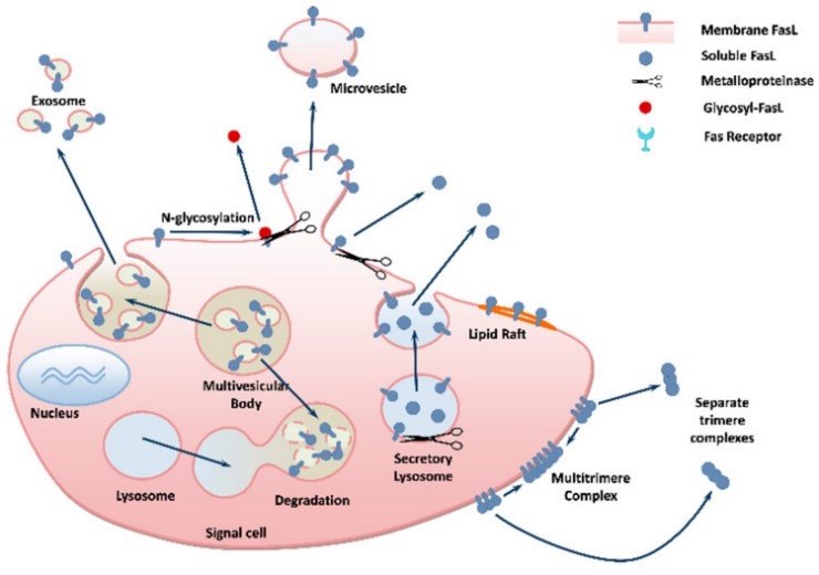
Fig1. Mechanisms of FasL secretion by cells. (Shulamit B Wallach-Dayan, 2021)
What is FASLG protein?
FASLG (Fas ligand) gene is a protein coding gene which situated on the long arm of chromosome 1 at locus 1q24. This gene is a member of the tumor necrosis factor superfamily. The primary function of the encoded transmembrane protein is the induction of apoptosis triggered by binding to FAS. The FAS/FASLGG signaling pathway is essential for immune system regulation, including activation-induced cell death (AICD) of T cells and cytotoxic T lymphocyte induced cell death. It has also been implicated in the progression of several cancers. The FASLG protein is consisted of 281 amino acids and its molecular mass is approximately 31.5 kDa.
What is the function of FASLG protein?
FASLG, also known as CD95L or FasL, is a cytokine capable of inducing apoptosis. By binding to Fas receptors, it triggers signal transduction events within cells that lead to programmed death of target cells, a process essential for maintaining the balance of the immune system and removing damaged or abnormal cells. In addition to its role in immune regulation, FasL is also involved in the occurrence and development of a number of diseases, such as certain types of cancer and autoimmune diseases. FasL is expressed on a variety of cell types, including T cells, natural killer (NK) cells, monocytes, neutrophils, mammary epithelial cells, and vascular endothelial cells.
FASLG Related Signaling Pathway
The main signaling pathway involved in FASLG protein is FAS related apoptosis pathway. By binding to its receptor FAS, the FASLG protein initiates a series of signal transduction events that ultimately lead to apoptosis. This signaling pathway plays an important role in regulating immune response, cell clearance and homeostasis. FASLG protein also interacts with other signaling pathways, such as NF-κB signaling pathway and p53 signaling pathway. FASLG plays an important role in the immune system, regulating the function of immune cells such as T cells, B cells and NK cells.
FASLG Related Diseases
1. Autoimmune diseases: The FAS-FASLG pathway plays an important role in autoimmune diseases, such as rheumatoid arthritis, systemic lupus erythematosus, etc. 2. Tumors: The FAS-FASLG signaling pathway plays an important role in immune escape and drug resistance of tumors, and FASLG is abnormally expressed in certain types of cancer, such as melanoma, breast cancer, and lung cancer. 3. Infectious diseases: Certain viruses and bacteria evade clearance by the immune system by interfering with the FAS-FASLG signaling pathway, leading to the occurrence of infectious diseases.
4. Neurological diseases: The FAS-FASLG signaling pathway also plays an important role in the development and functional regulation of the nervous system and is associated with neurodegenerative diseases such as Alzheimer's disease. 5. Inflammatory diseases: FAS-FASLG pathway plays an important role in inflammatory response and apoptosis of immune cells, and abnormal expression can lead to inflammatory diseases such as inflammatory bowel disease.
Bioapplications of FASLG
Understanding the FASLG signaling pathway could provide new ideas for the treatment of cancer and other diseases. For example, researchers are exploring how to harness or block the FASLG signaling pathway to develop new anti-cancer therapies. In addition, soluble forms of FASLG proteins, such as soluble Fas ligands produced by cleavage of exomatrix metalloproteinase MMP-7, may also be used as part of a therapeutic tool.
Case Study
Case study 1: Xiaoting Jin, 2014
Dichlorodiphenoxytrichloroethane (DDT) is a known persistent organic pollutant and liver damage toxicant. However, there has been little emphasis on the mechanism underlying liver damage toxicity of DDT and the relevant effective inhibitors. Hence, the present study was conducted to explore the protective effects of vitamin C (VC) and vitamin E (VE) on the cytotoxicity of DDT in HL-7702 cells and elaborate the specific molecular mechanisms.
The results demonstrated that p,p'-DDT exposure at over 10 μM depleted cell viability of HL-7702 cells and led to cell apoptotic. p,p'-DDT treatment elevated the level of reactive oxygen species (ROS) generation, induced mitochondrial membrane potential, and released cytochrome c into the cytosol, with subsequent elevations of Bax and p53, along with suppression of Bcl-2. When the cells were exposed to the NF-κB inhibitor (PDTC), the up-regulated expression of FasL was attenuated. Taken together, these findings provide novel experimental evidences supporting that VC or/and VE could reduce p,p'-DDT-induced cytotoxicity of HL-7702 cells via the ROS-mediated mitochondrial pathway and NF-κB/FasL pathway.
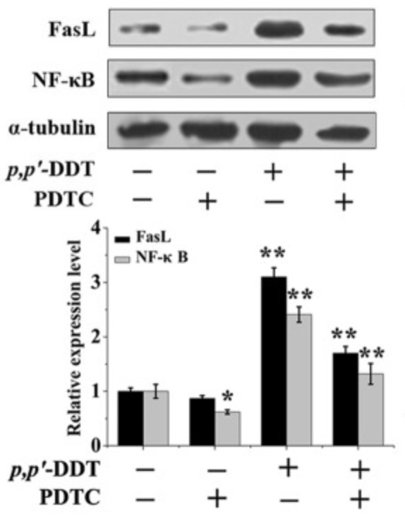
Fig1. p,p′-DDT induced the expression of FasL via activating NF-κB p65.

Case study 2: Mei Li, 2013
This study was conducted to analyze the molecular mechanisms responsible for anti-proliferation effects of glaucocalyxin A in cultured MCF-7 and Hs578T breast cancer cells. The concentration that reduced cell viability to 50% (IC50) after 72 h treatment was derived and potential molecular mechanisms of anti-proliferation using the IC50 were investigated as changes in cell cycle arrest and apoptosis. Gene and protein expression changes related to apoptosis were investigated by semi-quantitative RT-PCR and western blotting, respectively. Involvement of phosphorylated mitogen-activated protein kinases and JNK signaling in regulation of these molecules was characterized by western blotting. Cell viability decreased in a concentration-dependent manner and the IC50 was determined as 1 μM in MCF-7 and 4 μM in Hs578T cell. The results showed that the GLA-induced MCF-7 and Hst578T cell death was due to cell cycle arrest at the G2/M transition and was associated with activation of the c-jun N-terminal kinase (JNK) pathway.

Fig3. RT-PCR analysis of the expression of mRNA for FasL, p53, caspase 3 and β-actin in MCF-7 human breast cancers.

Quality Guarantee
High Purity
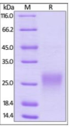
Fig1. SDS-PAGE (FASLG-46H) (PROTOCOL for western blot)
.
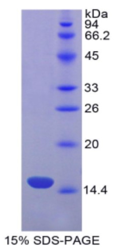
Fig2. SDS-PAGE (FASLG-624H) (PROTOCOL for western blot)
Involved Pathway
Fasl involved in several pathways and played different roles in them. We selected most pathways Fasl participated on our site, such as MAPK signaling pathway,Ras signaling pathway,Cytokine-cytokine receptor interaction, which may be useful for your reference. Also, other proteins which involved in the same pathway with Fasl were listed below. Creative BioMart supplied nearly all the proteins listed, you can search them on our site.
| Pathway Name | Pathway Related Protein |
|---|---|
| Autoi | TSHB,CD86,HLA-DPB1,HLA-DPA1,H2-AA,HLA-G,HLA-DRA,HLA-DQA1,IFNA21,IFNA7 |
| Allograft rejection | FASLG,HLA-DOA,IL12A,CD40LG,IL10,HLA-DQA1,GZMB,HLA-DPA1,HLA-DRB5,HLA-DOB |
| Natural killer cell mediated cytotoxicity | PIK3R5,KIR3DL1,PLCG2,CD247,RAET1E,ULBP2,KLRA8,RAC3,NFATC1,MICB |
| African trypanosomiasis | ICAM1,HBB,HBB-B1,GNAQ,SELE,VCAM1,PLCB4,IL10,FAS,HBB-B2 |
| Measles | EIF2AK3,IFNA4,STAT5A,IRF7,OAS1B,IL13,IRAK4,TRAF6,CCNE1,PIK3CA |
| ne thyroid disease | HLA-DOB,CD80,Ifna15,HLA-DRB5,Ifna11,CD40,HLA-DOA,H2-Q10,HLA-E,FAS |
| Ras signaling pathway | FGF6,IGF1R,PLCG1,GRB2,IKBKG,ANGPT2,MET,CALM4,RGL2,PLA2G6 |
| MAPK signaling pathway | MAPK1,CACNB1,GADD45BA,Fgf15,CACNB2,CACNA1SA,NTRK2,RASA1,PPP3CA,GNG12A |
| FoxO signaling pathway | IGF1RA,RAF1A,G6PC2,CDKN2B,BRAF,STK4,INS2,FOXO4,INSB,INSRB |

Fig1. Mechanisms of FasL action on "target" cells. (Shulamit B Wallach-Dayan, 2021)
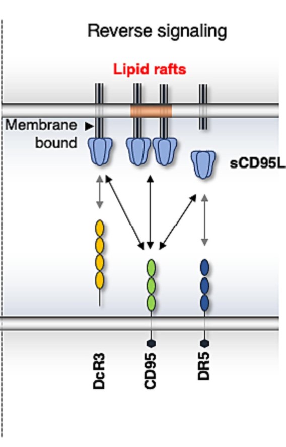
Fig2. CD95L aggregation can stimulate CD95 and induce different signaling pathways in the CD95L-expressing cell. (Layla Haymour, 2023)
Protein Function
Fasl has several biochemical functions, for example, cytokine activity,death receptor binding,tumor necrosis factor receptor binding. Some of the functions are cooperated with other proteins, some of the functions could acted by Fasl itself. We selected most functions Fasl had, and list some proteins which have the same functions with Fasl. You can find most of the proteins on our site.
| Function | Related Protein |
|---|---|
| cytokine activity | IL1B,GDF15,IFNB,CD40LG,LIF,FAM3D,MSTNB,IFNA16,CER1,BMP7B |
| death receptor binding | CASP8,BID,FASLG,MYD88,TMBIM1,FEM1B,FADD,NOL3,Casp3,TNFSF15 |
| tumor necrosis factor receptor binding | TNFSF10L3,EDA,TNFSF8,CD40LG,TRAP1,TNFSF15,TNF,TNFSF10,LTA,BRE |
Interacting Protein
Fasl has direct interactions with proteins and molecules. Those interactions were detected by several methods such as yeast two hybrid, co-IP, pull-down and so on. We selected proteins and molecules interacted with Fasl here. Most of them are supplied by our site. Hope this information will be useful for your research of Fasl.
Pstpip1;Ptpn12
Resources
Related Services
Related Products
References
- Zhu, J; Zhang, J; et al. Cumulus cells accelerate oocyte aging by releasing soluble Fas Ligand in mice. SCIENTIFIC REPORTS 5:-(2015).
- Liu, W; Lin, YT; et al. Hepatitis B virus core protein inhibits Fas-mediated apoptosis of hepatoma cells via regulation of mFas/FasL and sFas expression. FASEB JOURNAL 29:1113-1123(2015).

