Uncategorized Thursday, 2024/08/29
The selective metastasis of cancer cells to specific organs is a complex process that is influenced not only by anatomical factors, but also by biological and organ-specific microenvironmental factors. The pre-metastatic niche refers to the soluble factors and extracellular vesicles (EVs) produced by cells at the site of the primary tumor, which can alter the microenvironment of distant organs to accommodate migrating cancer cells and promote their growth. In addition, disseminated tumor cells (DTCs) can enter a dormant state in circulation and new tissue environments, evade immune surveillance, and interact with the tissue microenvironment to awaken from the dormant state. Finally, metastatic colonization requires multiple biological processes that depend on the inherent characteristics of cancer cells and the permissible tumor microenvironment provided by target organ cells.
In the process of liver metastasis of colorectal cancer, the inherent characteristics of colorectal cancer cells and the interactions between molecules and cells in the microenvironment can affect metastasis. Once CRC cells successfully enter the liver microenvironment, they interact with cell adhesion molecules on the sinusoidal endothelial cells, leading to the encapsulation and adhesion of disseminated tumor cells (DTCs).
When a large number of tumor cells invade the liver, the anti-tumor function of Kupffer cells (KCs) may be inhibited. Colorectal cancer cells may interact with KCs to stimulate the secretion of growth factors such as hepatocyte growth factor (HGF) and vascular endothelial growth factor (VEGF), which promote tumor cell proliferation and angiogenesis. Liver cells also play an important role in liver metastasis, and can release various growth factors that can promote tumor growth, motility, and invasion. However, the mechanism by which liver cells promote tumor metastasis is not fully understood.
Our Related Proteins

AFDN is located on chromosomes and is the main component of adhesion junctions (AJs), which form adhesion junctions between adjacent epithelial cells. AFDN, as a scaffold protein for adhesive connections, can recruit cadherin or linker proteins and interact with actin to promote the formation of adhesive connections. The role of AFDN in tumors has been reported for the first time in acute myeloid leukemia. In acute myeloid leukemia, fusion with lysine-specific methyltransferase 2A (MLL) leads to changes in AFDN subcellular localization and activation of the Ras signaling pathway, promoting the progression of acute myeloid leukemia and resulting in poor prognosis. However, AFDN mainly exerts tumor-suppressive effects in various solid tumors.
Loss or down-regulation of AFDN expression is associated with poor prognosis of colorectal cancer, osteosarcoma, endometrial cancer, breast cancer, and pancreatic cancer. In pancreatic cancer, the loss of AFDN not only destroys adhesion and tight junction, but also up-regulates the expression of Snail, promoting epithelial-mesenchymal transformation (EMT) and metastasis. AFDN also maintains adhesive connections and inhibits tumor cell migration and invasion by interacting with cystic fibrosis transmembrane conductance regulator (CFTR) in CRC. However, the biological function and molecular mechanism of AFDN in the liver of colorectal cancer patients are still unclear.
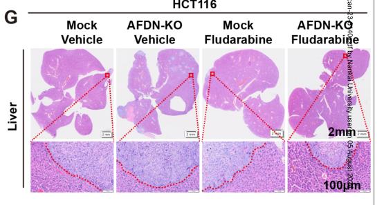
Recently, researchers from the Fourth Affiliated Hospital of Zhejiang University School of Medicine published an article titled ” AFDN deficiency promotes liver tropism of metastatic colorectal cancer ” in the journal Cancer Res. The study showed that AFDN deficiency can promote liver metastasis in colorectal cancer, providing a new perspective and understanding for studying the mechanism of liver metastasis in colorectal cancer.
Liver metastasis is the main cause of morbidity and mortality in patients with colorectal cancer. A better understanding of the biological mechanisms of liver tropism and metastasis in colorectal cancer can help determine better prevention and treatment strategies.
In this study, researchers conducted genomic CRISPR functional loss screening in a mouse colorectal cancer model and found a deficiency in AFDN, a protein involved in establishing and maintaining cell-cell contacts and a driving factor for liver metastasis. Elevated expression of AFDN is associated with prolonged survival in colorectal cancer patients. Colorectal cancer cells lacking AFDN preferentially metastasize to the liver rather than the lungs. The absence of AFDN in the primary site of colorectal cancer cells promotes cancer cell migration and invasion by disrupting tight intercellular connections.
In addition, through the JAK-STAT signaling pathway, CXCR4 expression is increased in colorectal cancer cells with afdn deficiency, thereby reducing the motility of afdn deficient colorectal cancer cells and promoting their colonization in the liver.
In summary, the role of AFDN in cancer progression is very complex. AFDN cannot reduce intercellular TJs, thereby promoting the migration and invasion of colorectal cancer cells. AFDN deficiency can also inhibit their movement in the liver, ultimately leading to the formation of liver metastases. However, the specific and hepatotropic molecular mechanisms of AFDN deficiency in colorectal cancer metastasis still need to be elucidated. This study provides a new perspective and understanding for investigating the mechanism of liver metastasis in colorectal cancer, however, further research is needed.
Related Products and Services
Protein Expression and Purification Services
Reference
Liao S, Deng J, Deng M, Chen C, Han F, Ye K, Wu C, Pan L, Lai M, Tang Z, Zhang H. AFDN deficiency promotes liver tropism of metastatic colorectal cancer. Cancer Res. 2024 Jul 24. doi: 10.1158/0008-5472.CAN-23-3140. Epub ahead of print. PMID: 39047222.